
Facilities
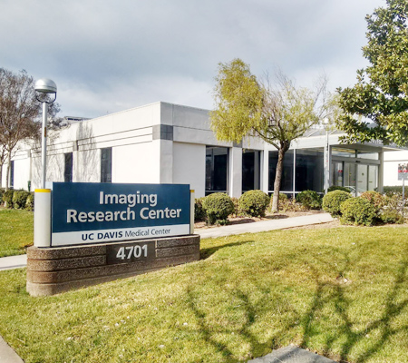
Sacramento
The University of California, Davis, established the Imaging Research Center (IRC) in 1998 to support imaging science research and to promote the use of modern imaging methods in basic science and clinical investigations of the brain and body.
The IRC is located in a 13,000 sq. ft. building on the UC Davis Health campus in Sacramento, and currently houses a research-dedicated whole-body 3T Siemens Prisma MRI System. A wide range of imaging studies is performed at the IRC. The IRC supports basic science and clinical research that investigates the structure and function of the nervous system, including perceptual, motor, and cognitive function, using real-time functional imaging techniques, as well as research that investigates systemic physiology and morphology in health and disease.
The IRC is dedicated to research involving advanced imaging and image processing technologies and serves UC Davis Health and the Main Campus. The Center houses a whole-body 3T MRI system and general imaging research support equipment. Imaging physics faculty and technical support personnel have offices at the IRC and continually improve the research infrastructure and support faculty research projects.
Siemens Prisma Scanner
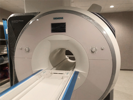
The IRC operates a 3T Prisma whole-body MRI system (Siemens Medical Solutions, Erlangen, Germany), which is fully equipped for advanced brain imaging. This system has a short-bore (2 m) magnet, a fast-gradient system that enables high-speed structural and functional imaging, and a 64-channel data acquisition system for parallel imaging.
The Prisma combines a maximum gradient amplitude of 80 mT/m with a 200 T/m/s slew rate, enabling higher SNR across all regions of the body. Newer and more powerful gradient systems are critical for fast imaging techniques such as Echo Planar Imaging (EPI), the modality of choice for both fMRI and DWI, which are used extensively by UC Davis investigators. For EPI sequences used in fMRI, signal loss due to underlying B0 inhomogeneity, for example, in the orbital frontal cortex (OFC), is greatly reduced by reducing the effective echo time (TE), echo spacing (ESP), and by increasing the k-space acquisition matrix size, using parallel imaging.
The Prisma uses a parallel transmit system with two independent coils. This means that the B1 field can be homogenized by varying the relative phase between the two transmit coil elements. This is referred to as B1-shim. B1 homogeneity is essential for obtaining signal uniformity across the field of view for neuroimaging while speeding up specific absorption rate-limited scans.
The Prisma offers an improved Radio Frequency (RF) transmit and receive system with digital input and output, enhancing signal quality by transmitting the RF signal over fiber-optic cables. This eliminates electromagnetic crosstalk between different RF channels and interference from spurious RF sources, improving the signal-to-noise ratio (SNR). Another benefit resulting from increased spatial and temporal SNR is reduced total exam time. This allows studies involving children or patients with anxiety to get higher-quality data in less time.
Additionally, dedicated force compensation on each axis yields low vibrations and acoustic noise, while high-performance cooling for each axis enables full duty cycle over long-term measurements with outstanding stability. As a result, the gradients of the Prisma system create significantly less noise pressure than in previous systems. Many participants, particularly children and patients with anxiety disorders, complain about the acoustic noise of the scanner.
Reduction in acoustic noise is expected to substantially increase the percentage of adults and children who can complete their scanning sessions. Three multi-channel RF head coils are available: the Siemens 64-channel brain coil that takes full advantage of the 64 receiver channels and provides the highest signal-to-noise ratio, a 32-channel coil that provides continuity with ongoing studies acquired with the previous TIM Trio system, and a 16-channel coil which provides excellent SNR while also providing more space between the head and the coil for headphones and goggles for enhanced auditory and visual presentation and simultaneous EEG/fMRI studies.