Residency Program - Case of the Month
September 2018 - Presented by Trevor Starnes, M.D.
Clinical History
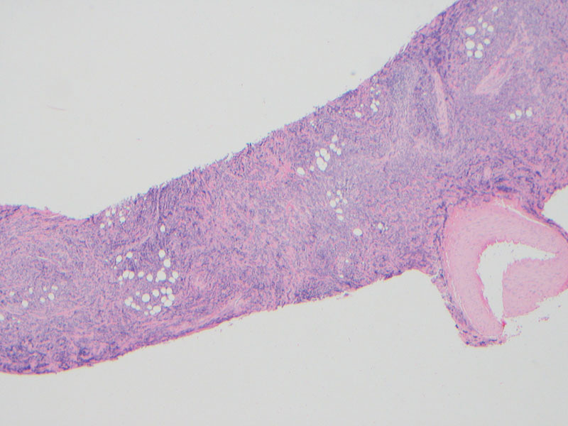
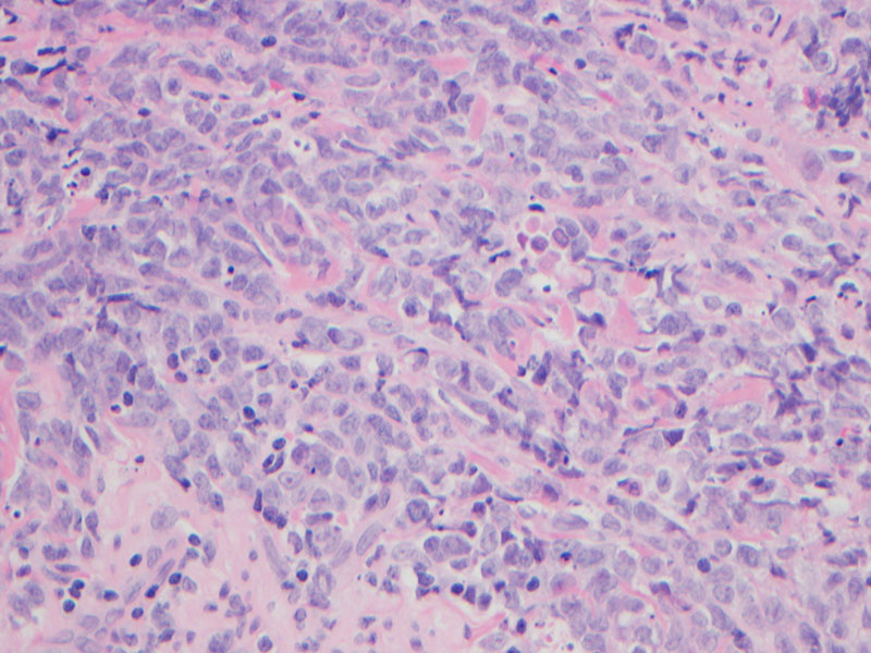
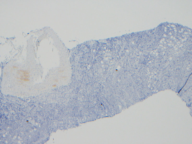
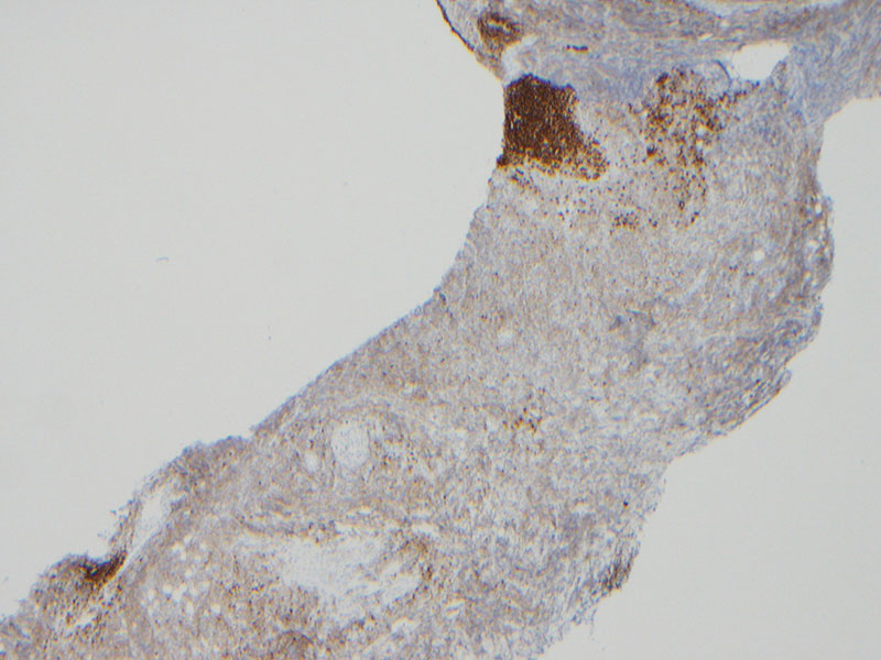
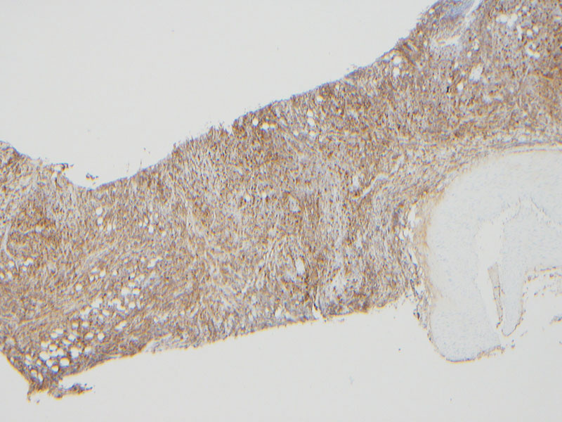
A previously healthy adolescent male presents with one week of jaundice and abdominal pain. Liver enzymes are elevated, and a CBC is normal. Radiology imaging reveals an obstructing mass at the pancreatic head. A biopsy of the mass is obtained. Bone marrow biopsies and peripheral blood flow cytometry (not shown) are normal.
Click on images to enlarge.
Which is the most likely diagnosis?
Choose one answer and submit.


 Meet our Residency Program Director
