Residency Program - Case of the Month
March 2014 - Presented by Rebecca Sonu, M.D.
Clinical History:
The patient is a 47 year-old male with no significant past medical history who presented with a palpable mass of several years in the anterior and lateral aspects of the left lower leg. The mass was first attributed to a musculoskeletal injury and no further evaluation was obtained. However, the lesion continued to progress in size with severe pain. A surgical biopsy was performed yielding a pathologic diagnosis. At that time, a bone survey showed metastatic lytic lesions in the left iliac crest, lumbar and thoracic spine. Given the presence of metastatic disease, the patient was given systemic chemotherapy for six months. Radiation therapy was also administered for the lesions in the spine.
A post-chemotherapy MRI of the lower extremity showed a large soft tissue mass (30 x 6 x 6 cm) with low, intermediate and high areas of T2 signal intensity. There was heterogeneous mild to moderate enhancement scattered throughout the lesion. The mass extended into the deep posterior and peroneal compartments and extended superiorly adjacent to the iliotibial band insertion of the tibia. Several areas of fibular erosion and intramedullary extension were present in the proximal one third of the fibula (Figure 1). After treatment, the patient was taken to surgery for a radical resection of the left lower fibula.
 |
| Figure 1 |
Gross Images:
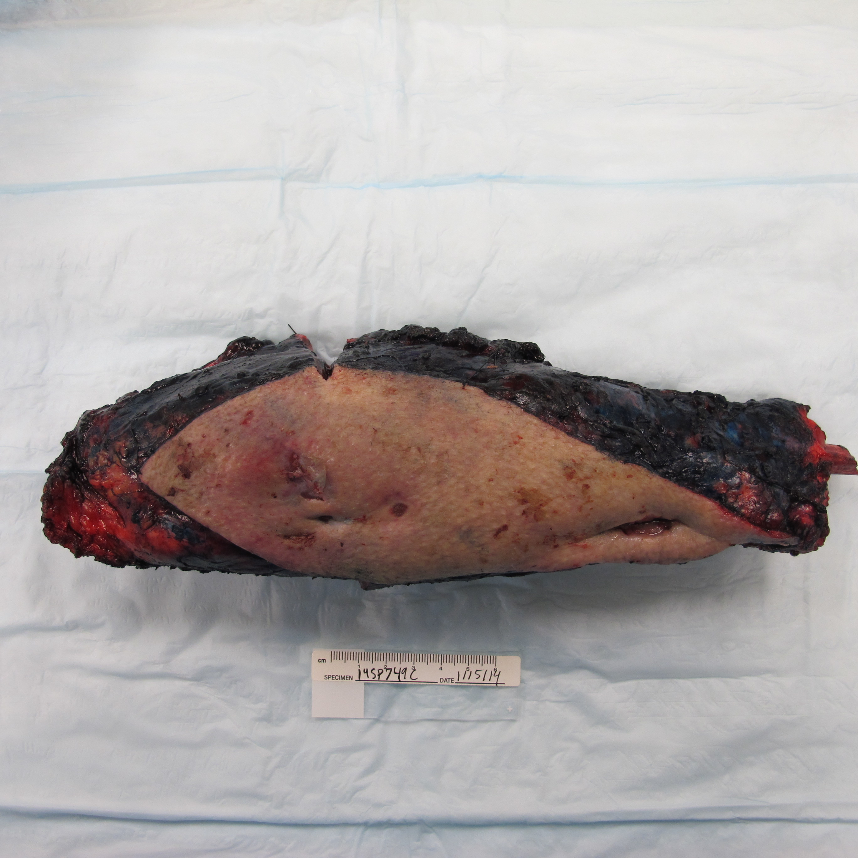
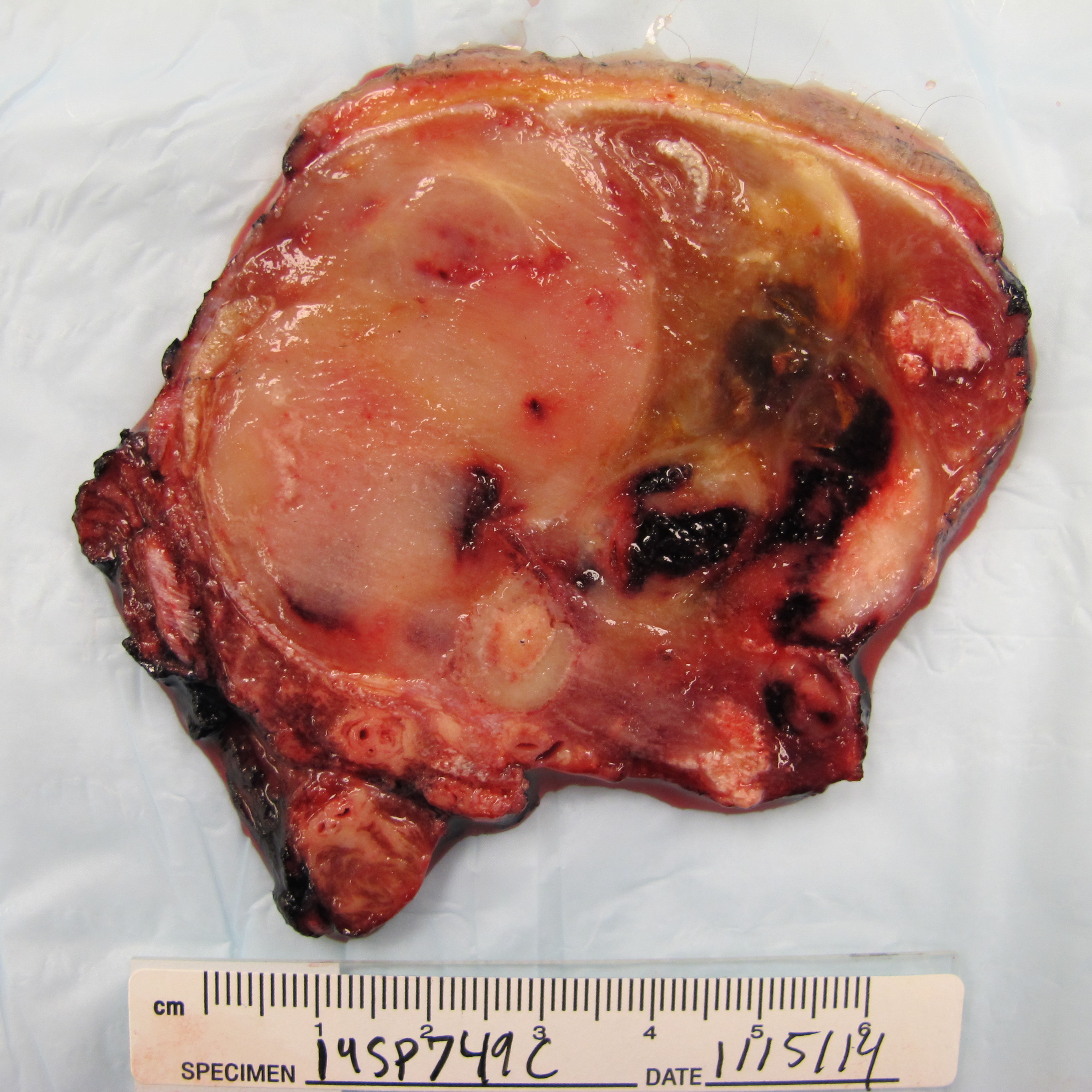
Received fresh was a portion of fibula with surrounding soft tissue and an attached tan skin ellipse measuring 27.6 cm (superior to inferior) x 7.5 cm (medial to lateral) x 5.4 cm (anterior to posterior) (Figure 2). The skin ellipse (22.1 x 7.1 cm) contained multiple red-pink to red-tan lesions on the skin surface. The specimen was frozen and then thinly sectioned to reveal an ill-defined (20.5 (superior to inferior) x 6.5 (anterior to posterior) x 6.2 cm (medial to lateral) white-pink focally hemorrhagic mass (Figure 3) extending to within 0.2 cm from the closest lateral margin of resection. The mass grossly appeared to encase the fibula and abut the deep margin of the resection. A portion of the tumor was submitted for molecular studies.
 |
 |
|
| Figure 2 | Figure 3 |
Microscopic Images:
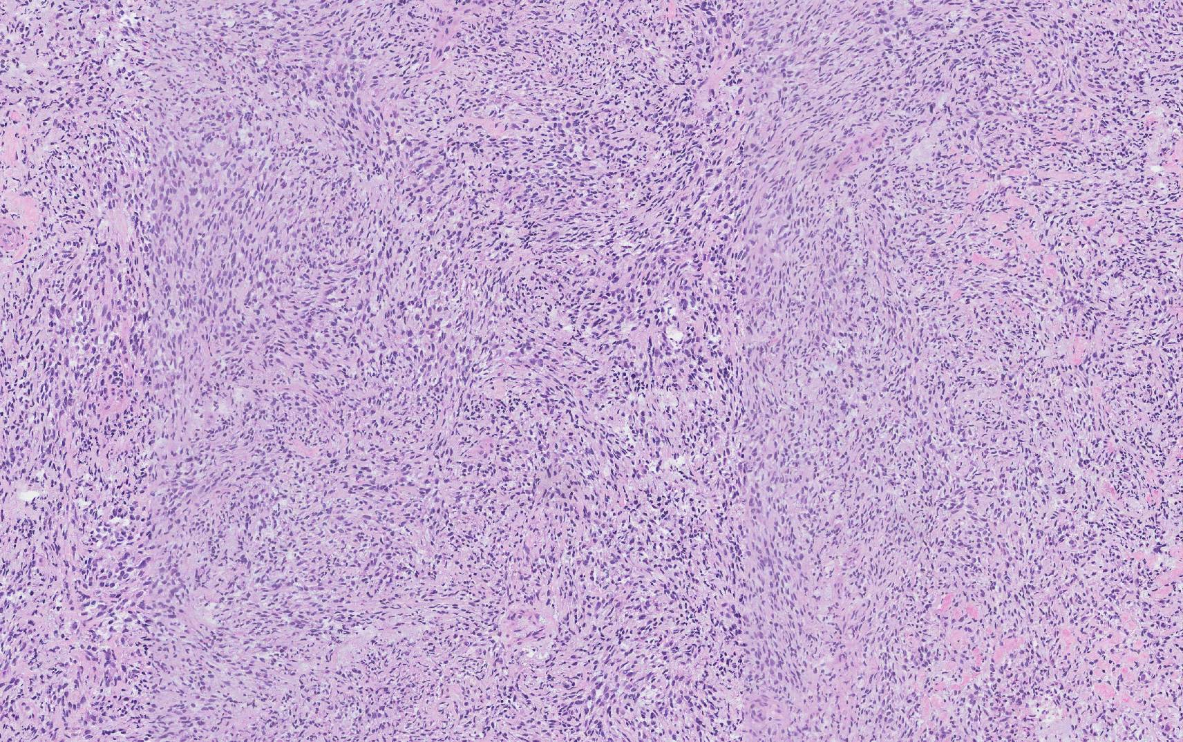
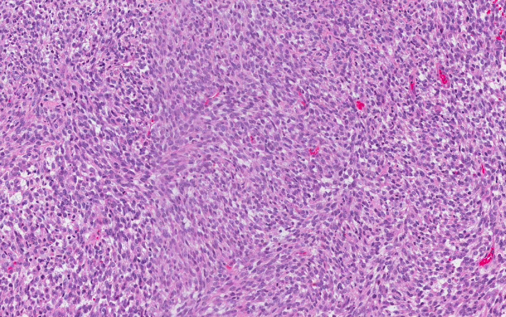
Microscopically, the areas of residual tumor consisted of a monotonous population of spindled cells in a whorled-like pattern with areas of fasciculation. (Figure 4). The cells had a fibroblast-like appearance with plump and elongated nuclei with coarse chromatin and eosinophilic cytoplasm (Figure 5 and 6). Rare mitotic figures were present. Scattered areas of dense pink hyalinization were present throughout. Post-treatment necrosis was abundant. Glandular or heterologous elements were not identified.
 |
 |
 |
||
| Figure 4 | Figure 5 | Figure 6 |
Immunohistochemistry Results (from prior biopsy):
Vimentin: Positive in tumor cells
Epithelial membrane antigen: Positive in tumor cells
Desmin: Negative in tumor cells
Pan-cytokeratin: Scattered positive in tumor cells
Cytogenetics Results:
Out of 20 cells, 65% of the cells showed some variation from cell to cell:
59-63<3n>,YY,t(X;18)(p11.2;q11.2),+der(X)t(X;18),1,add(3)(p11),dup(8)(q24.1q11.2),-9,-11,-13,-13,-14,-15,-18,+21,+0-3mar[cp13]/46,XY[7]
- A translocation between chromosomes X and 18 plus an extra copy of the derivative chromosome X generated from the X;18 translocation
- loss from <3n> of chromosomes 1,9,11,13,14,15 and 18
- gain from <3n> of chromosome 21
- added material on the short arm of chromosome 3
- a chromosome 8 with inverted duplication of the segment 8q11.2-8q24.1
- gain of a variable number (0-3) of marker chromosomes.
In 35% of the cells, a normal male chromosome complement was seen.

 Meet our Residency Program Director
