December 2023 – Presented by Dr. Lorene Chung (Mentored by Dr. Frank Melgoza)
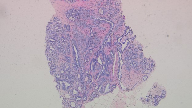
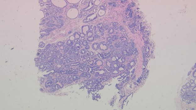
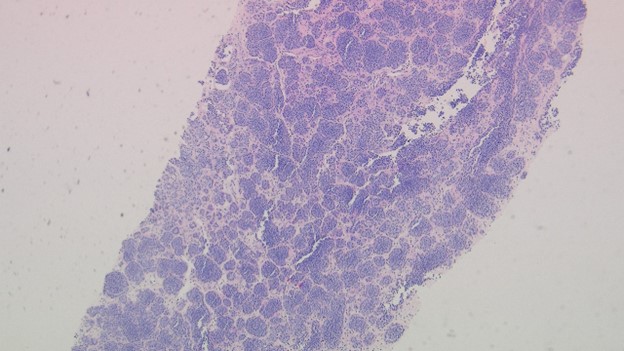
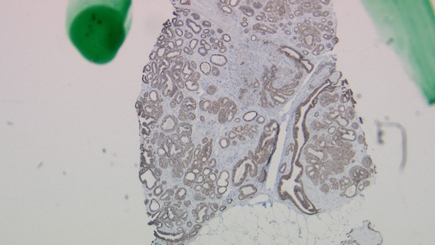
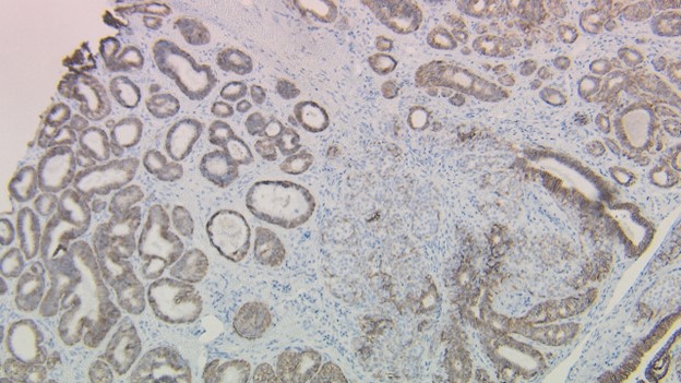
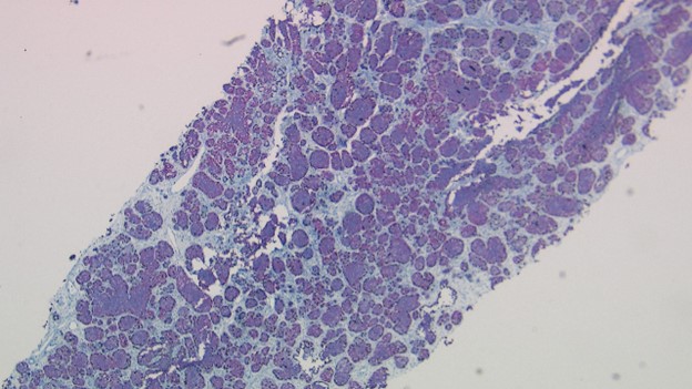
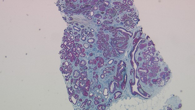
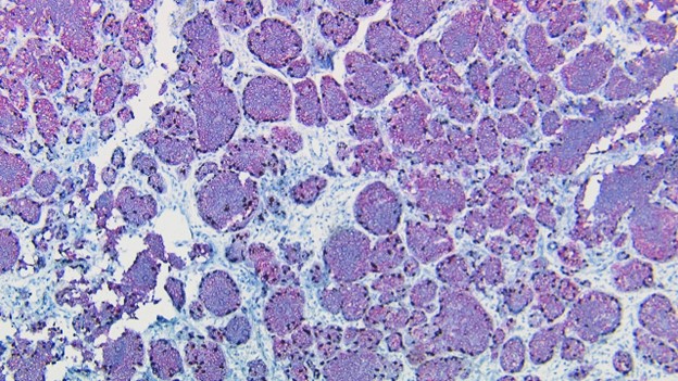
A 52-year-old, postmenopausal G1P1 female with a family history of breast carcinoma presents for routine mammogram and is found to have multiple left breast lesions. Menarche was at 14 years old. Imaging finds a 1.6 x 1.6 cm high density, partially circumscribed mass with microcalcifications located at 3:00, 6cm posterior to the nipple and a 1.4 cm mass at 12:00, 9cm posterior to the nipple line. Core needle biopsy is shown below. What is the most likely diagnosis?
Atypical lobular hyperplasia (ALH)
Ductal carcinoma in situ (DCIS)
Lobular carcinoma in situ (LCIS)
Usual ductal hyperplasia (UDH)
Invasive lobular carcinoma (ILC)
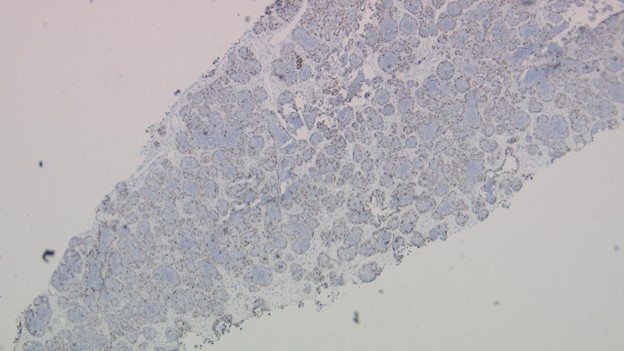
E-Cadherin
Atypical lobular hyperplasia (ALH)
Ductal carcinoma in situ (DCIS)
Lobular carcinoma in situ (LCIS)
Usual ductal hyperplasia (UDH)
Invasive lobular carcinoma (ILC)

 Meet our Residency Program Director
