
August 2020 – Presented by Ying Liu (Mentored by Mingyu Cheng)
Clinical History
A 67-year-old man with worsening bilateral otalgia, dry cough, hemoptysis, fatigue and hoarseness for the past 2 months. CT scan of the neck demonstrated an infiltrative mass measuring 3.2 x 2.1 x 1.2 cm (Figure 1). The mass involved bilateral vocal cords and anterior commissure with extension into the infra glottic trachea with associated narrowing of the proximal tracheal. There is no invasion of the cartilages or anterior neck soft tissues. The patient underwent direct laryngoscopy, an ulcerative exophytic mass occupying more than 70% of the glottis was identified and excised (Figure 2).
Pathology Review
Pathological evaluation shows both subepithelial proliferation of spindle cells in haphazard fascicles (Figure 3-4), as well as poorly differentiated squamous cell carcinoma component with hyperchromatic, pleomorphic nuclei with eosinophilic cytoplasm (Figure 5-6). Frequent mitotic figures are noted. Immunohistochemical staining shows tumor cells are positive for AE1/AE3 and vimentin. Additional immunohistochemical stains are performed and the results are listed in Table 1.
Table 1
| ANTIBODY | RESULT |
| AE1AE3 | Positive |
| Vimentin | Positive |
| P40 | Positive |
| CK5/6 | Focal positive |
| P16 | Negative |
| S100 | Negative |
Images:
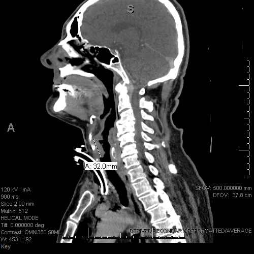
Figure 1: CT scan of the neck demonstrated an infiltrative 3.2 cm mass involving bilateral vocal cords with extension into the infra glottic trachea.
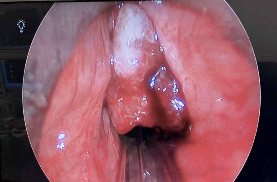
Figure 2: Laryngoscopy showed an ulcerated, exophytic mass occupying more than 70% of the glottis.
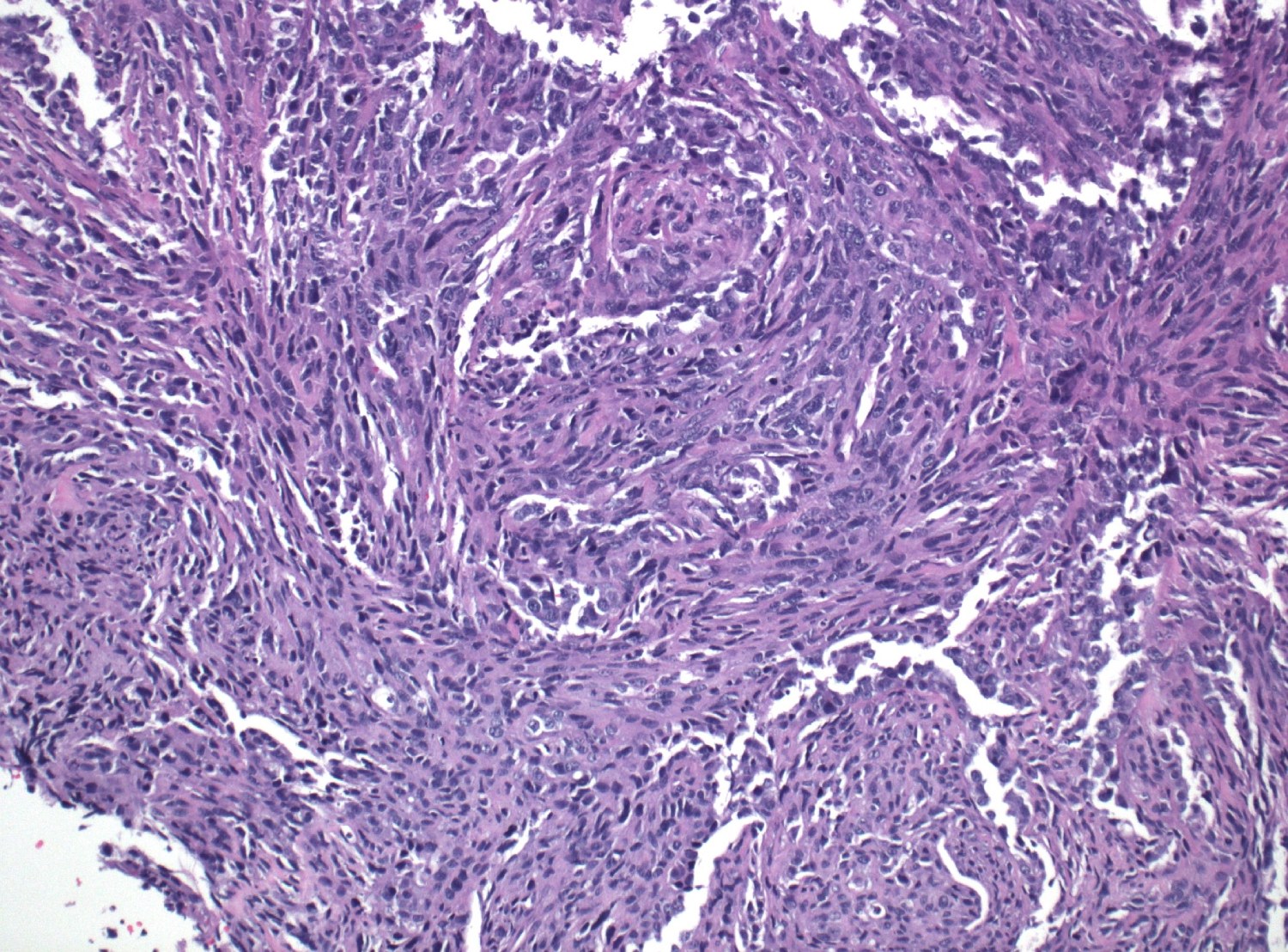
Figure 3: Spindle cells, low power
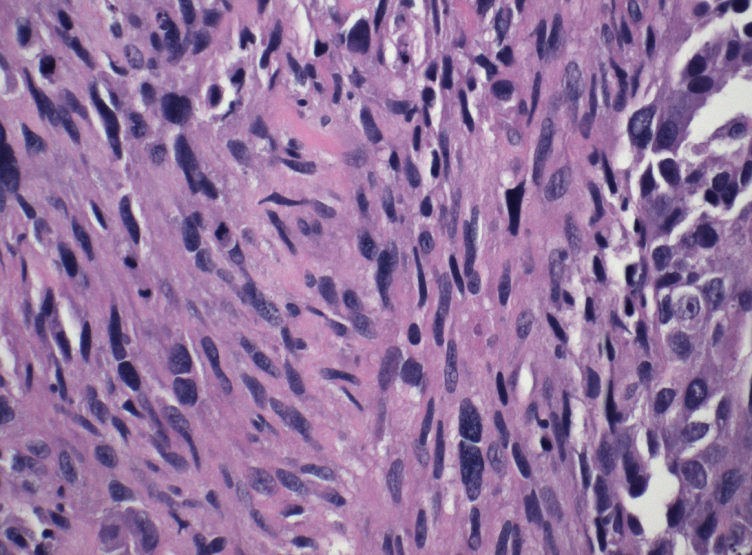
Figure 4: Spindle cells, high power
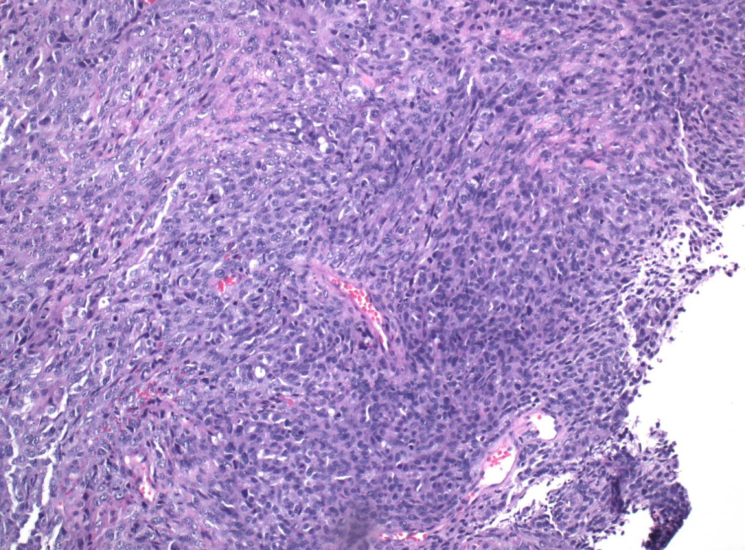
Figure 5: Poorly differentiated tumor cells, low power
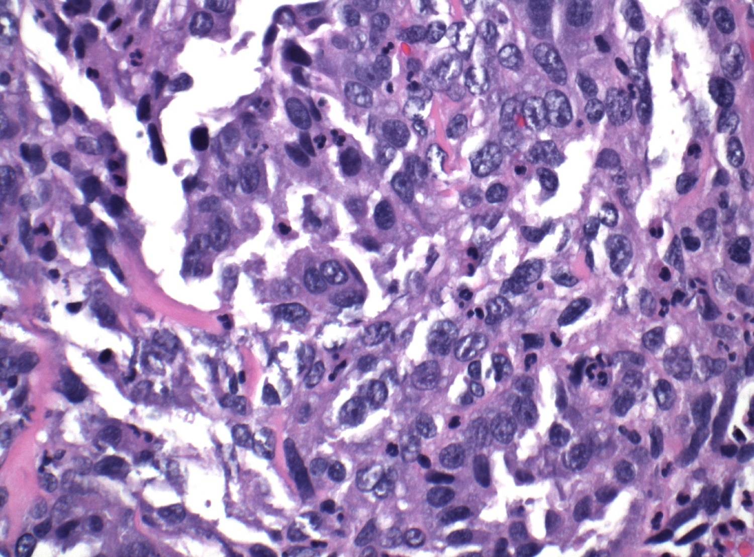
Figure 6: Poorly differentiated tumor cells, high power
 Meet our Residency Program Director
