Residency Program - Case of the Month
July 2018 - Presented by Dongguang Wei (Mentored by Tao Wang)
Clinical History
A 17-year-old female presented with right femur fracture and underwent intermedullary nailing and open reduction internal fixation. She complained right hip pain again 9 months after the surgery. X-ray revealed hardwire failure and non-union of the fracture. Biopsy demonstrated only vascular tissue. She underwent revision with hardware replacement. Culture of the non-union fracture comes back negative. She complained of persistent right hip pain, X-ray showed right femoral neck fracture around the nail and she was admitted again for a right femur removal of hardware and proximal femoral replacement.
Further Imaging Studies
CT and X-ray shows intraosseous hemangioma, diffuse demineralization/osteolysis of the proximal femur, underlying severe osteoporosis and tapering of bone.
Pathology Review
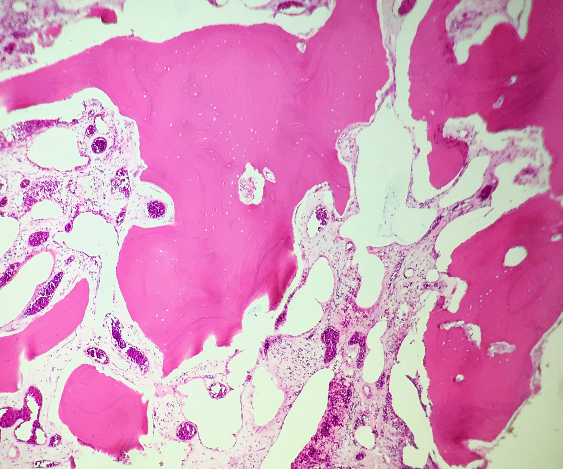
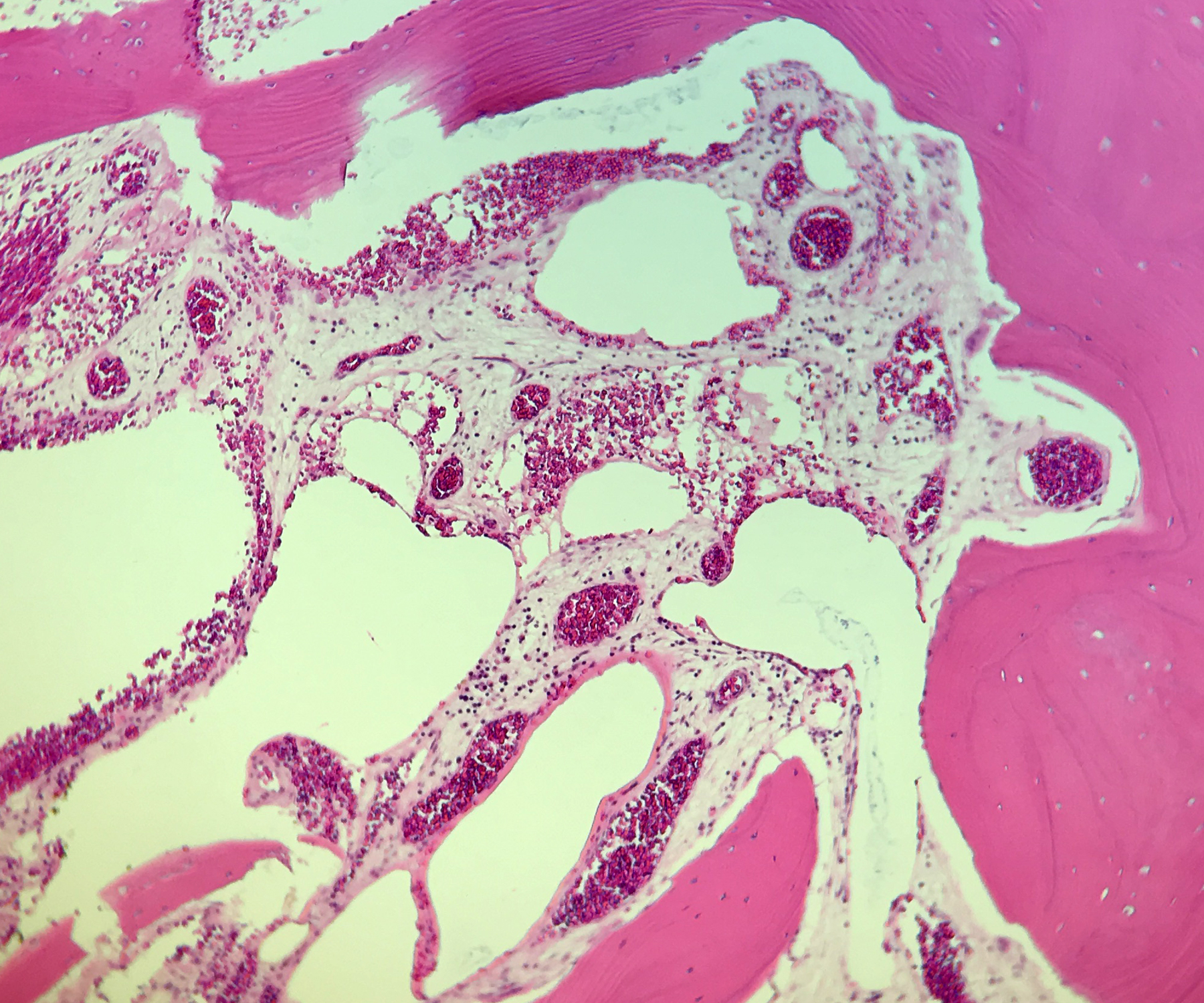
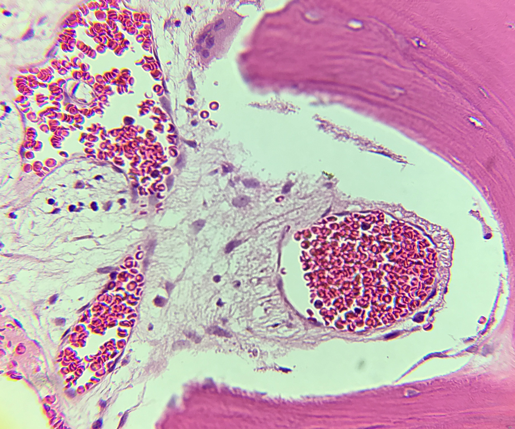
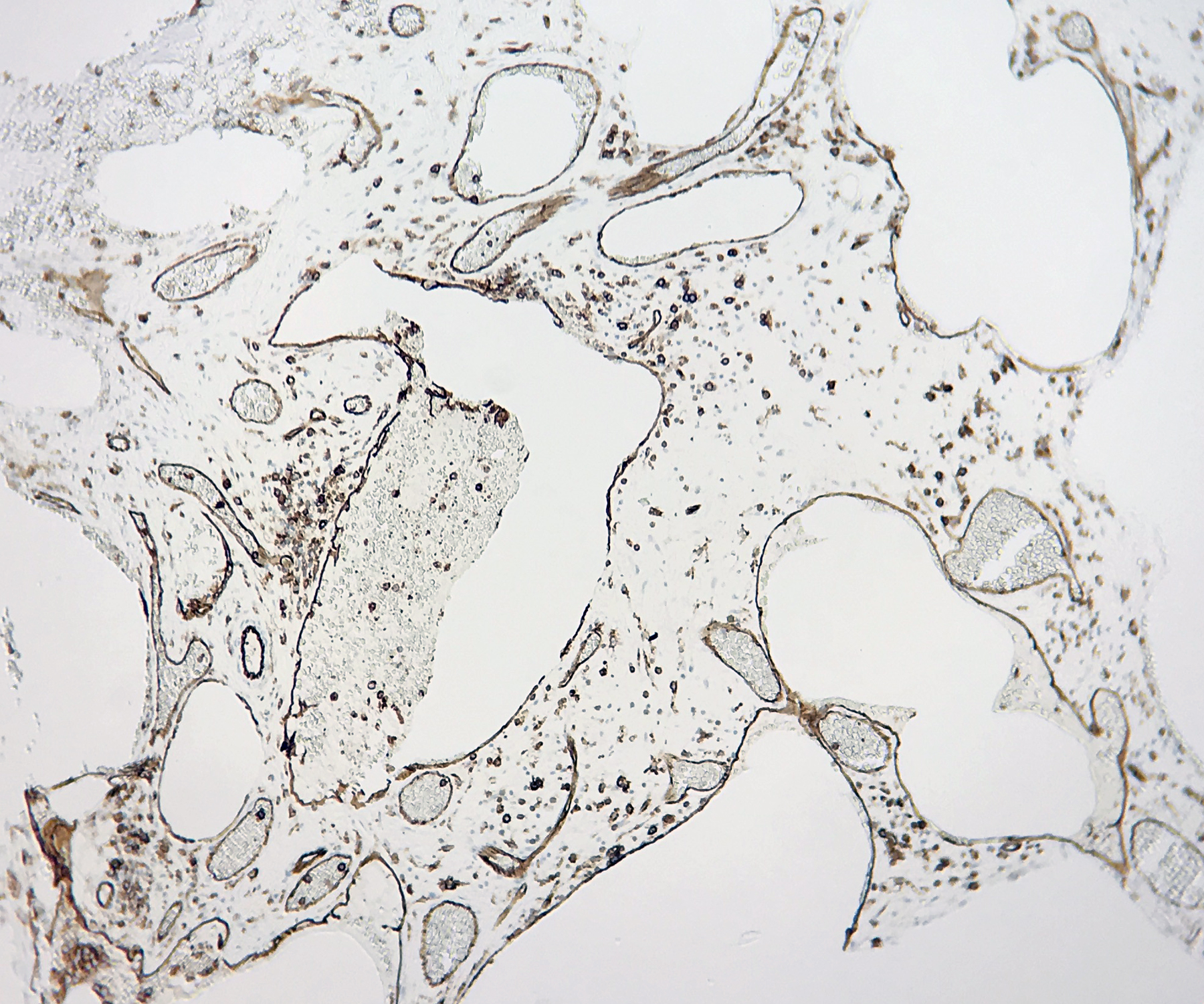
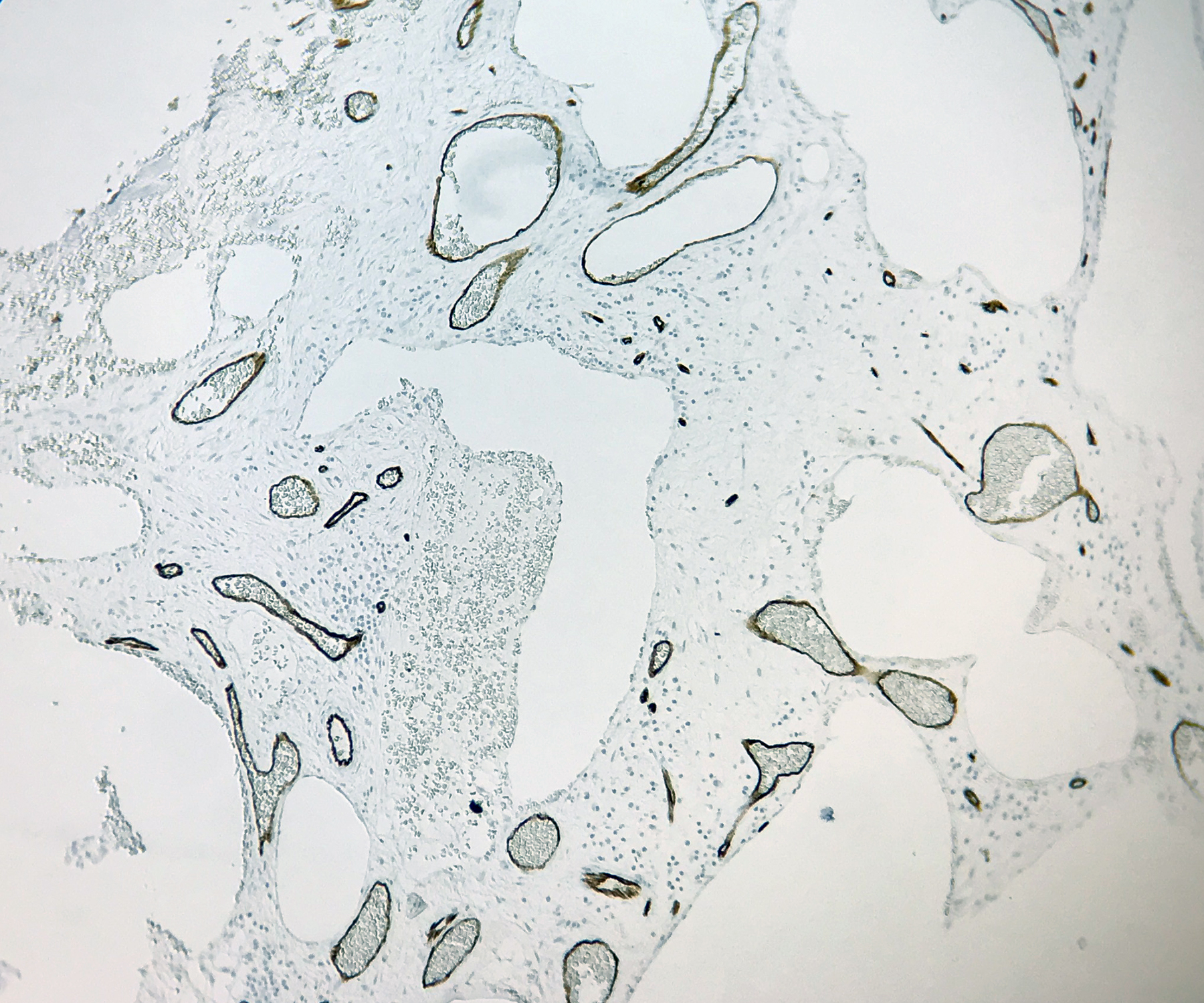
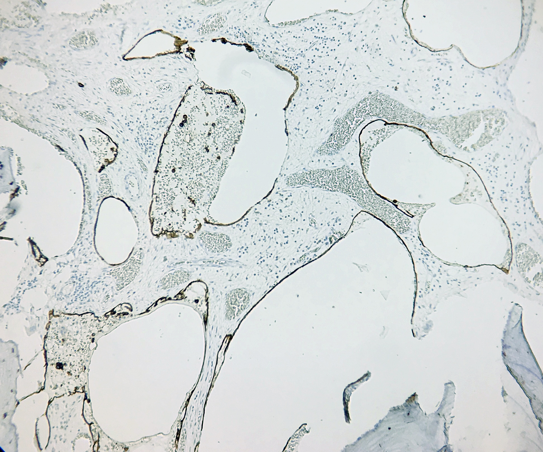
Multiple pathology reviews showed fragments of viable bones with remodeling changes, negative for acute inflammation; Benign lymphovascular proliferation with network of thin-walled vessels highlighted by CD31, CD34, and D2-40. Osteoclasts are noted within the adjacent bones, and demonstrate scalloped osteoclastic activity.
Images
Click on images to enlarge.
Which of the following is most likely the diagnosis?
Choose one answer and submit.
D. Gorham-Stout Syndrome (Vanishing Bone Disease)
> Learn more about this diagnosis.

 Meet our Residency Program Director
