
Residency Program - Case of the Month
September 2017 - Presented by Amir Ghorbani
Clinical History
A 5 week old female presented with blood in her diaper. On examination, she was unremarkable without any evidence of pain, fever, or rash. However, placing a Foley catheter revealed gross hematuria. She was admitted for further work up.
RADIOLOGIC FINDINGS
Abdominal ultrasound showed a 3.0 x 2.6 x 2.0 cm hypoechoic mass which appeared to be confined to the central portion of the right kidney (Figure 1).
Abdominal MRI showed a mass which was slightly hyperintense on T2 (Figure 2) and slightly hypointense on T1 (Figure 3). The mass was well-circumscribed and there was no evidence of hydronephrosis or infiltration of surrounding fat.
PATHOLOGIC FINDINGS
The patient underwent a right radical nephrectomy. Our pathology lab received a 32.0 g, 5.8 x 3.6 x 3.1 cm nephrectomy specimen. Sectioning revealed a well-demarcated 3.1 x 2.8 x 2.1 cm mass in the middle third of kidney with tan fleshy cut surface and areas of hemorrhage. It was directly beneath the renal capsule but appeared to be confined to the kidney. The remainder of the specimen was unremarkable. (Figure 4)
Microscopic examination showed a well-demarcated highly cellular mass composed of fascicles of plump spindle cells with numerous mitoses. (Figures 5, 6, 7)
FISH testing was positive for ETV6 (12p13.2) rearrangement.
Click image to enlarge.
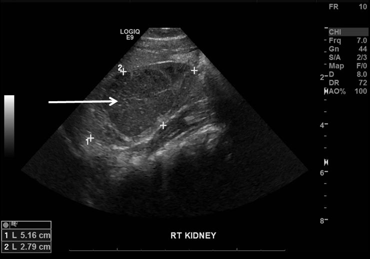
Figure 1: Ultrasound shows a hypoechoic mass in the right kidney (arrow).
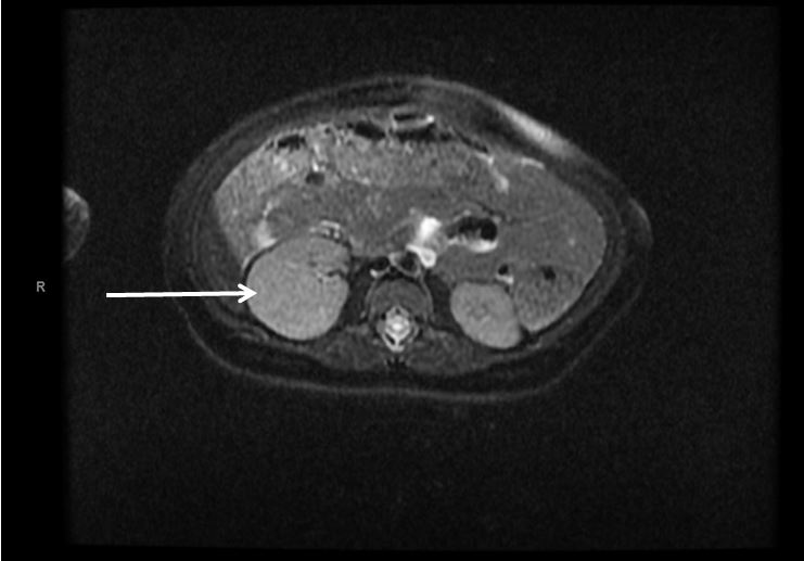
Figure 2: MRI-T2 shows a slightly hyperintense well-circumscribed mass in the right kidney (arrow).
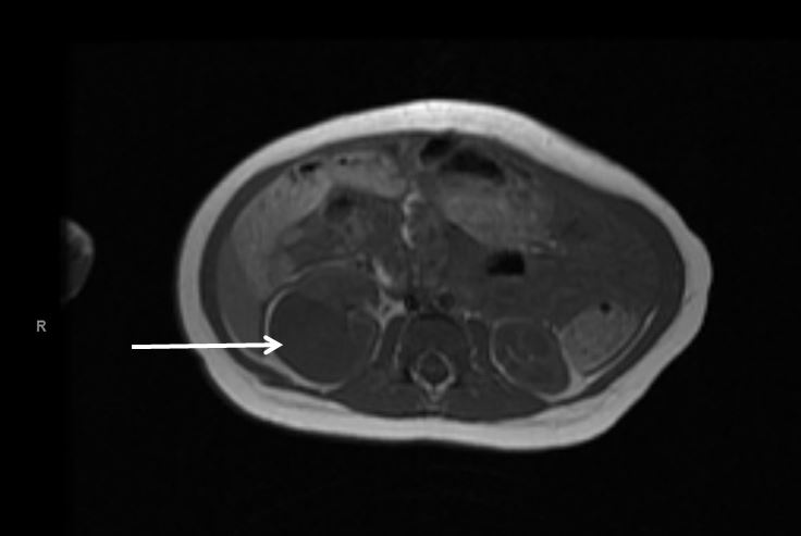
Figure 3: MRI-T1 shows a slightly hypointense right renal mass (arrow).
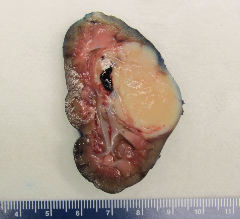
Figure 4: Well-circumscribed solid mass with fleshy tan cut surface and areas of hemorrhage.

Figure 5: Densely packed spindle cells with an area of hemorrhage.
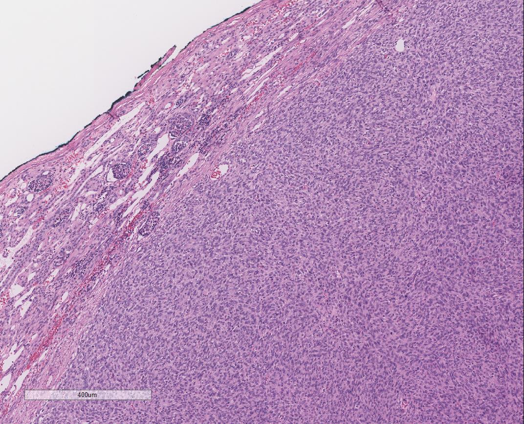
Figure 6: Tumor with “pushing” border, dense cellularity.
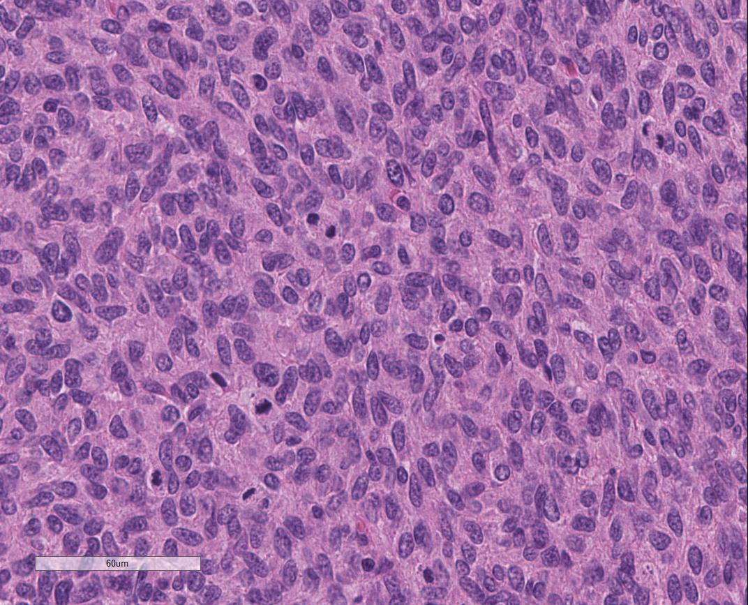
Figure 7: Densely packed plump spindle cells with high mitotic rate.
What is the diagnosis?
Choose one answer and submit.
 Meet our Residency Program Director
