
Residency Program - Case of the Month
April 2017 - Presented by Guofeng Gao and Regina Gandour-Edwards
Clinical History
A 74-year-old man has a 1.5 year history of a right inferior pole renal mass seen incidentally on CT scan. He has been asymptomatic and under observation with imaging studies. Surveillance CT revealed a slight increase in mass size from 1.8 cm to 2.2 cm. PET/CT scan demonstrated a FDG avid lesion. He is referred for biopsy and radiofrequency ablation of this right renal mass.
Microscopic Description
The CT guided core biopsy showed bland and uniform, oval or spindle cells arranged in parallel bundles, cuboidal cells in tightly packed tubules, and prominent vacuoles in a myxoid/mucinous stroma (Figure 1 and 2). The nuclei are round and uniform with evenly dispersed chromatin and prominent small single nucleoli. Mitoses are rare and no necrosis is present. IHC results: Tumor cells were Positive for CK7 (Figure 3), AMACR (Figure 4), RCC (Figure 5), PAX8 (Figure 6) and PAX2 (Figure 7); and Negative for CD10 (Figure 8).
Click on the images to enlarge
Figure 1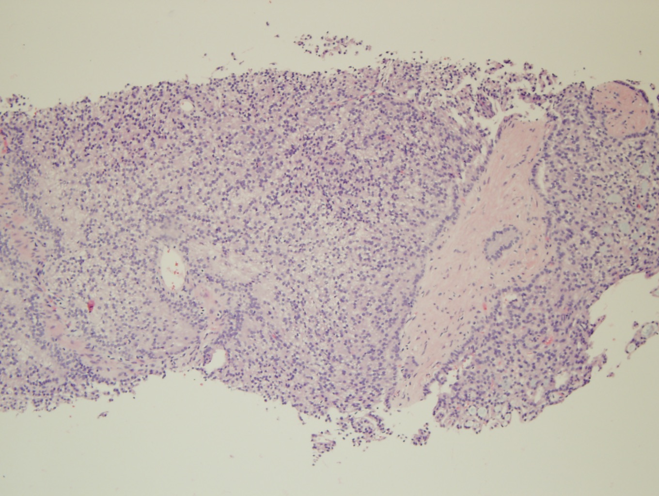
Figure 2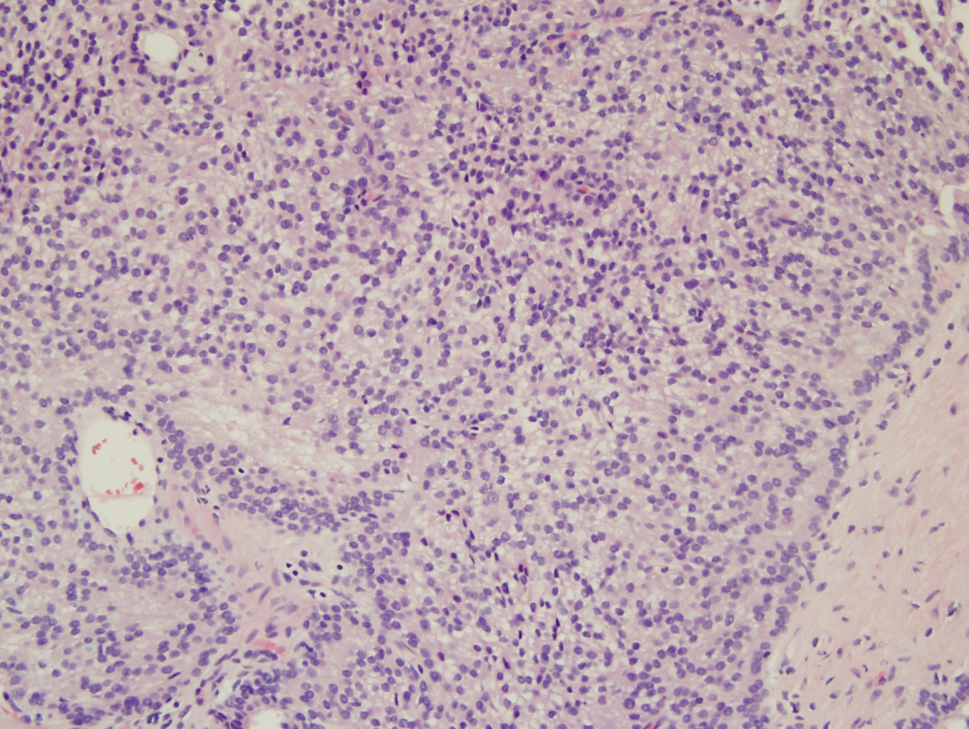
Figure 3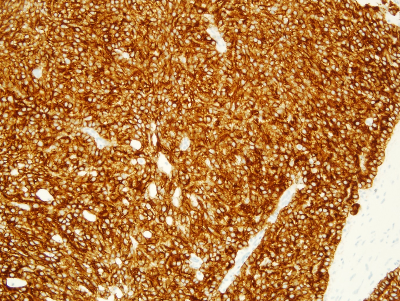
Figure 4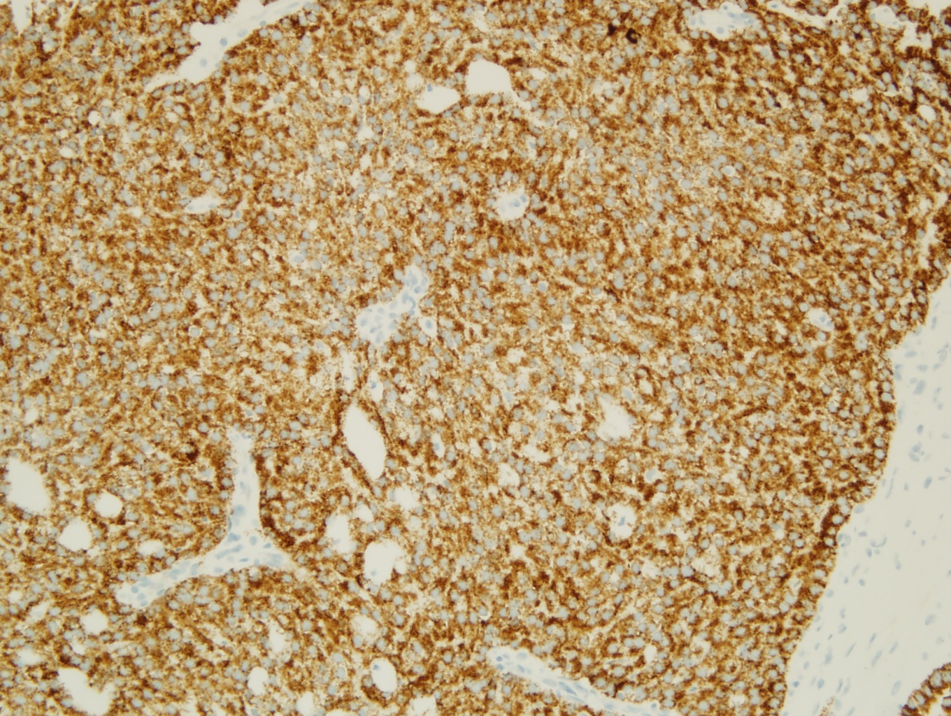
Figure 5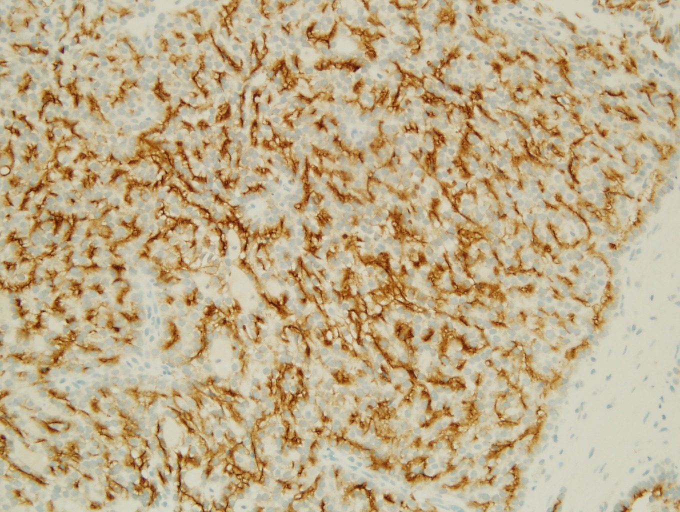
Figure 6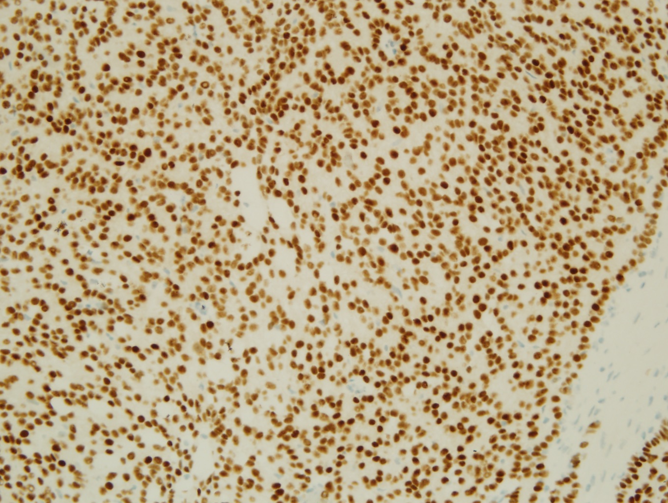
Figure 7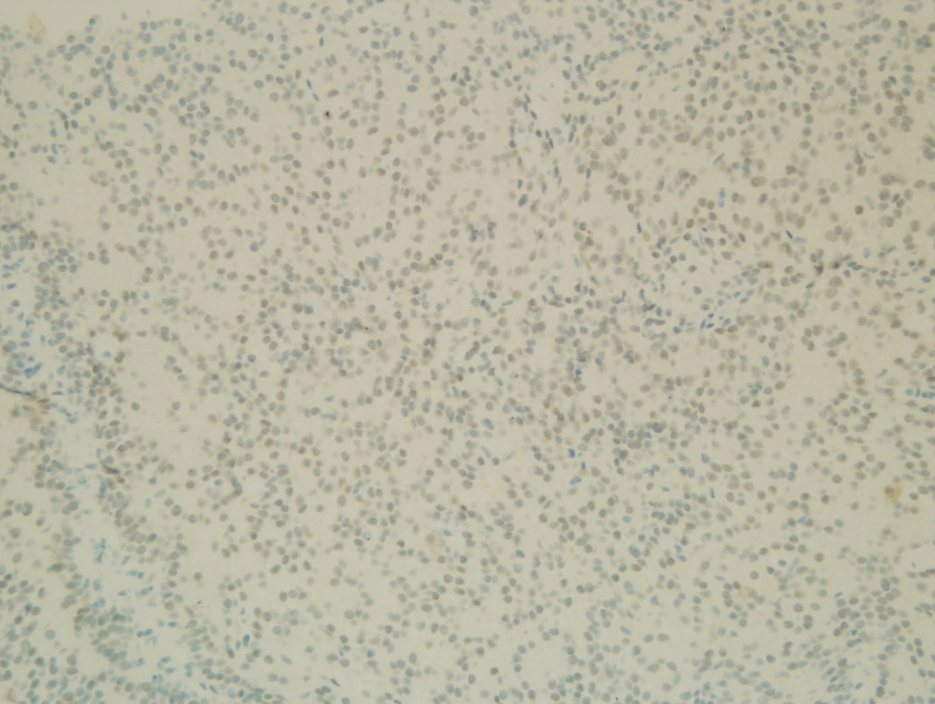
Figure 8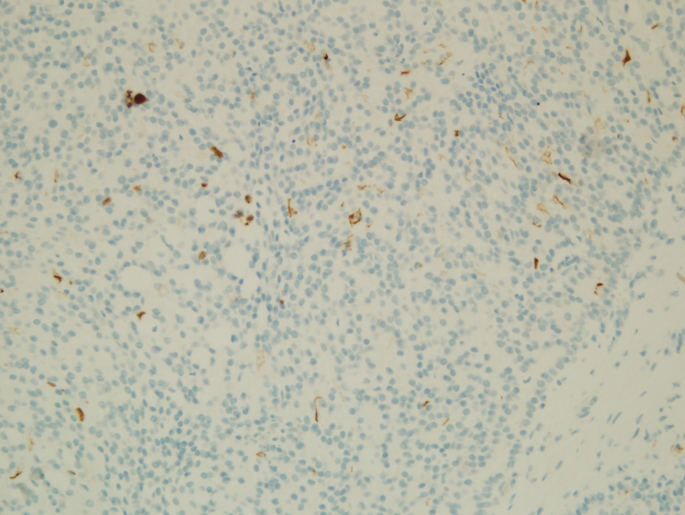
What is the diagnosis?
Choose one answer and submit.
E. Mucinous Tubular and Spindle Renal Cell Carcinoma (the name in the CAP protocol)
> Learn more about this diagnosis.
 Meet our Residency Program Director
