Residency Program - Case of the Month
October 2014 - Presented by John Rodrigo, M.D.
Clinical History:
The decedent is a 55-year-old Caucasian male with a past medical history of diffusely metastatic carcinoma, severe chronic obstructive pulmonary disease (on 4 L home oxygen therapy), extensive smoking history, and asbestos exposure history. He had symptomatic lesions and pathologic fractures at the lumbosacral spine, the left pelvis, the left femur and the left upper extremity. He was not a candidate for systemic treatment or surgery but was considering possible radiation therapy.
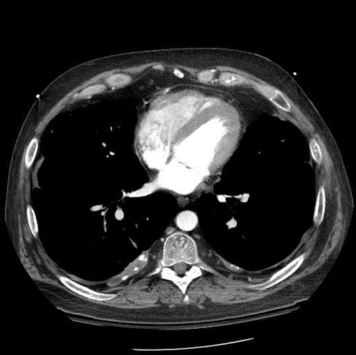
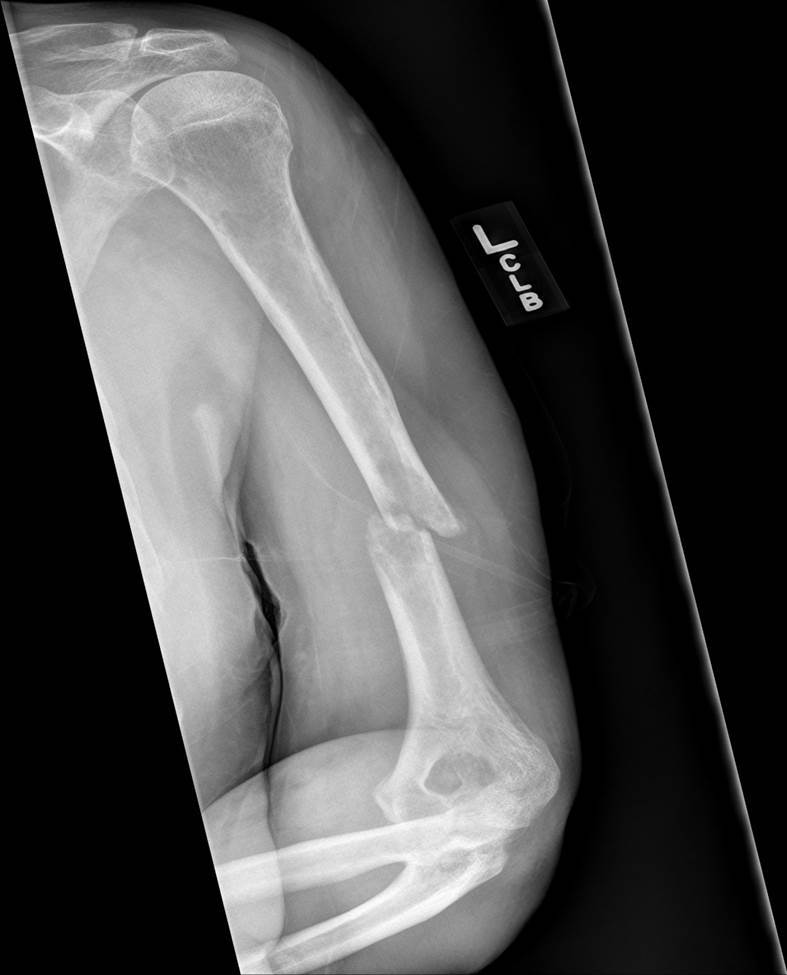
A CT of the chest revealed diffuse extensive pleural soft tissue masses consistent with metastasis throughout the left thorax associated with calcifications. A CT guided FNA of the right sacrum was performed revealing malignant cells. Further studies to help better classify the malignant cells could not be performed due to insufficient numbers of cells. An x-ray of the left humerus demonstrated a midshaft pathologic fracture with an anteromedial displacement of the distal fracture fragment. A PET-CT scan performed revealed bilateral pleural effusions. The left was greater than the right and showed malignant studding and calcifications. Multiple lytic osseous lesions were seen. The decedent had elected for hospice care and was pronounced dead soon thereafter.
 |
 |
|
| CT of Chest | X-Ray of Left Humerus |
Gross:
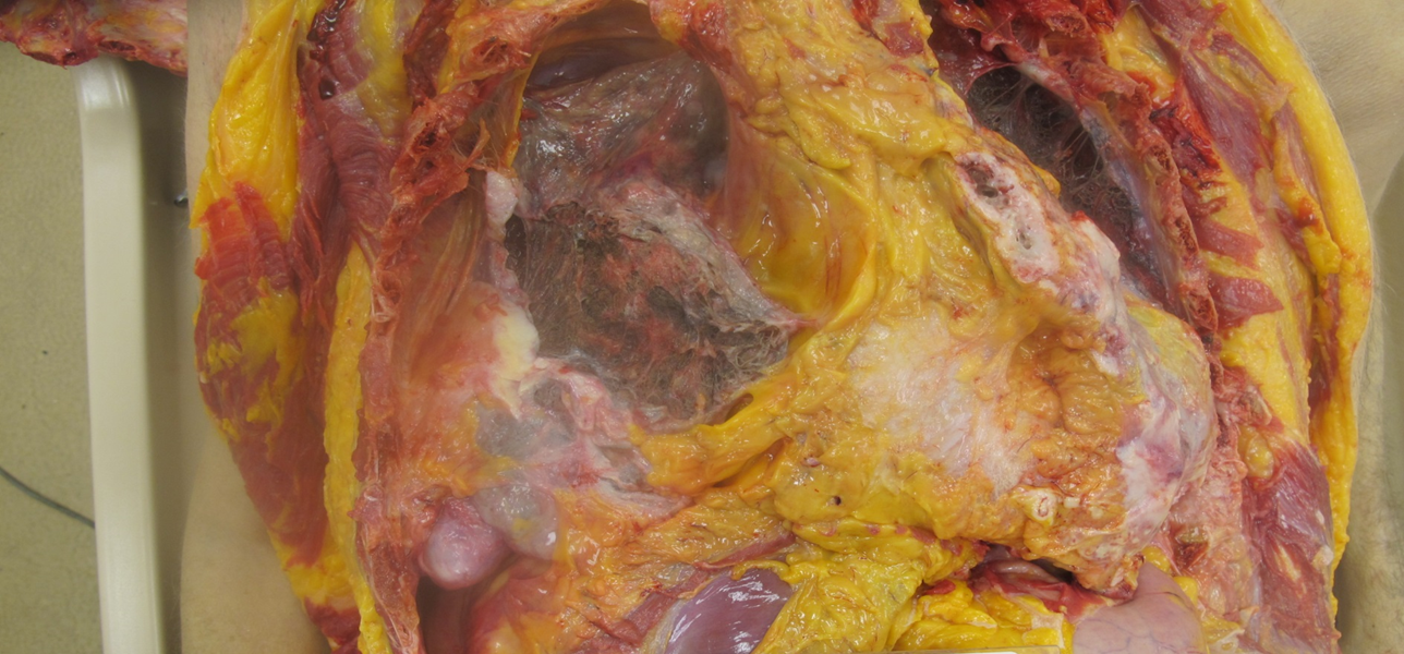
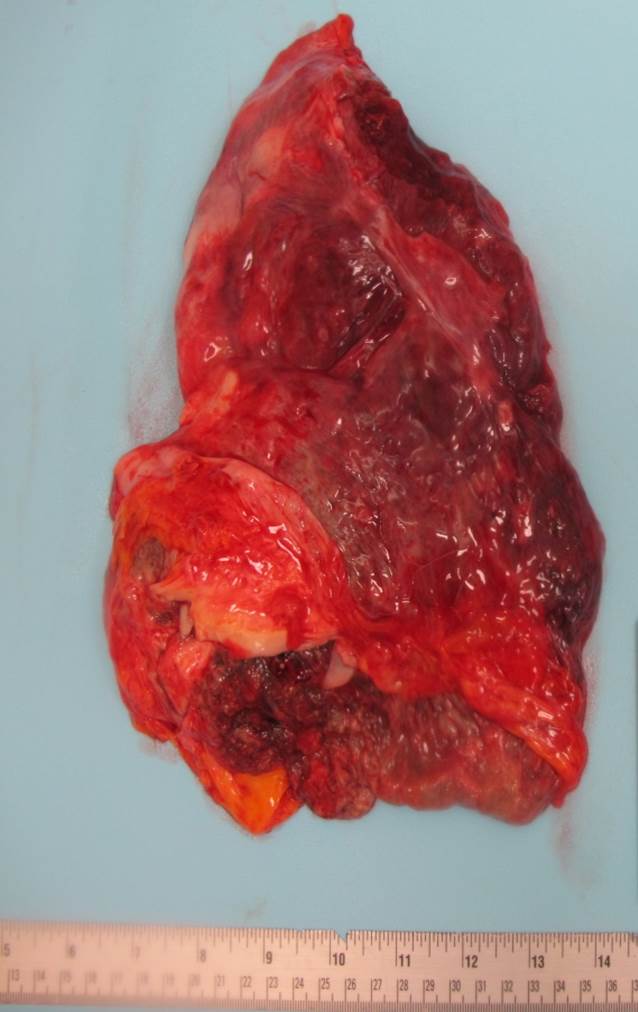
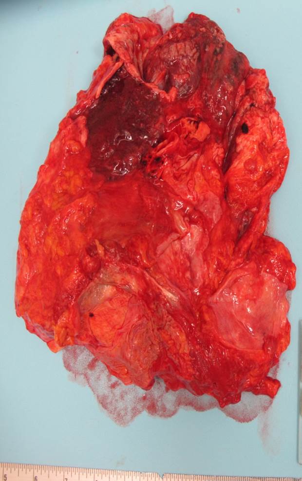
On autopsy, the pericardial sac was encased in a firm, tan white, honey-combed soft tissue that extends to the left lateral thoracic wall. Both lungs were almost double their normal weights. The left lung had multiple adhesions to the left thoracic wall and multiple firm white nodules, the largest of which measured 6.7cm x 5.5cm x 4.6cm. The posterior medial right lung has a single white firm plaque that is adherent to the lung wall, it measured 5.5 x 2.6 cm. There was a tan brown firm nodule measuring 0.4 x 0.2 x 0.2 cm located in the omental fat. Several of the vertebrae in the lumbosacral area were grossly involved by tumor.
 |
 |
 |
||
| Pericardial Sac | Left Lung | Right Lung |
Immunohistochemistry:
Calretinin: negative in cells of interest
CK7: diffusely intracellular positive in cells of interest
CK20: negative in cells of interest
TTF-1: negative in cells of interest
Special Stains:
Iron stain: positive in alveolar macrophages
Mucicarmine: scattered focal areas of intracellular mucin present
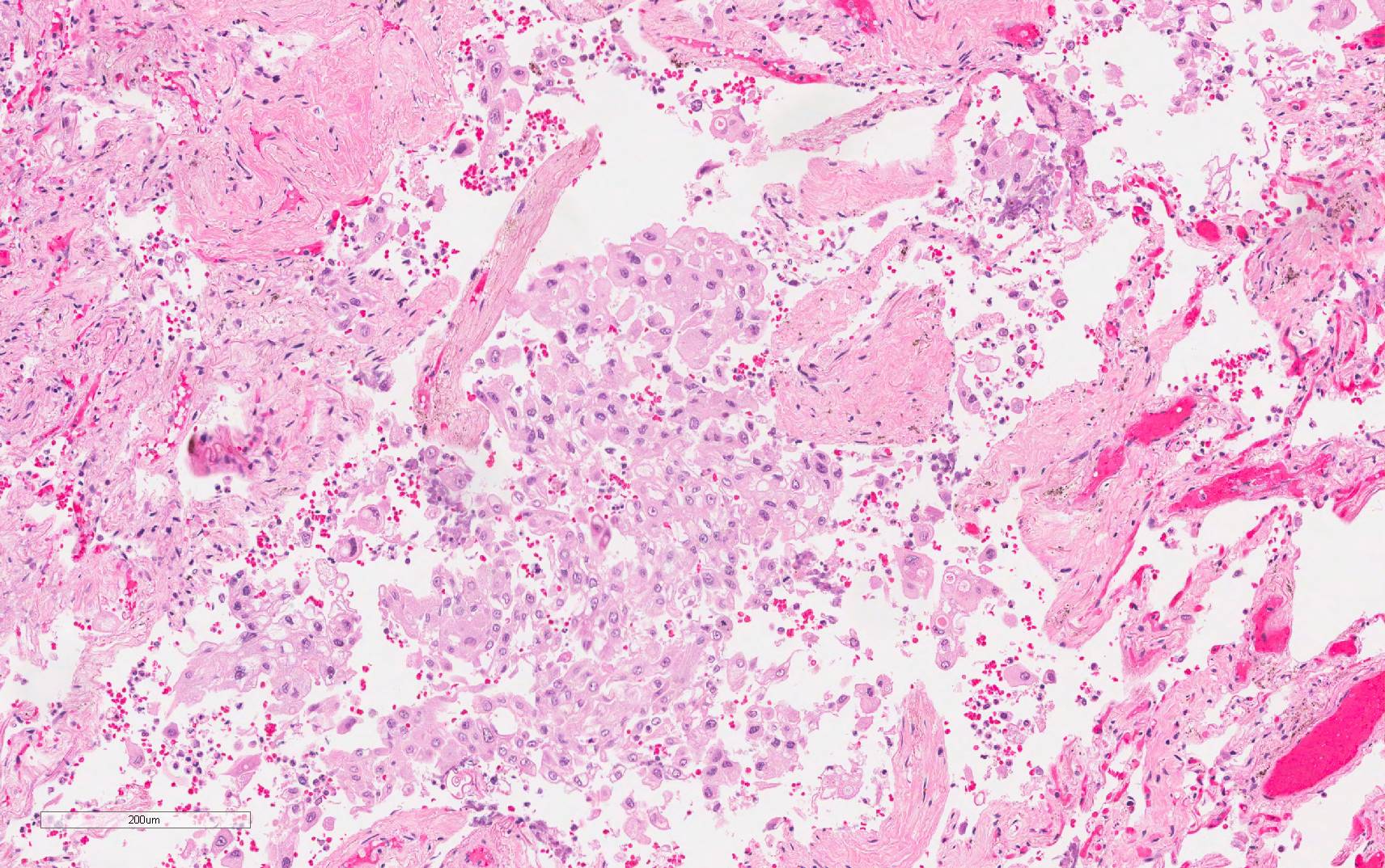
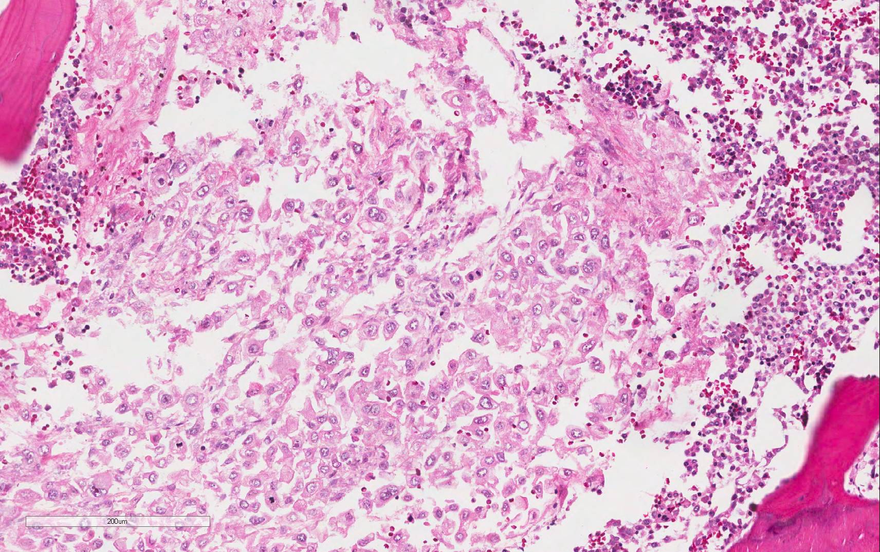
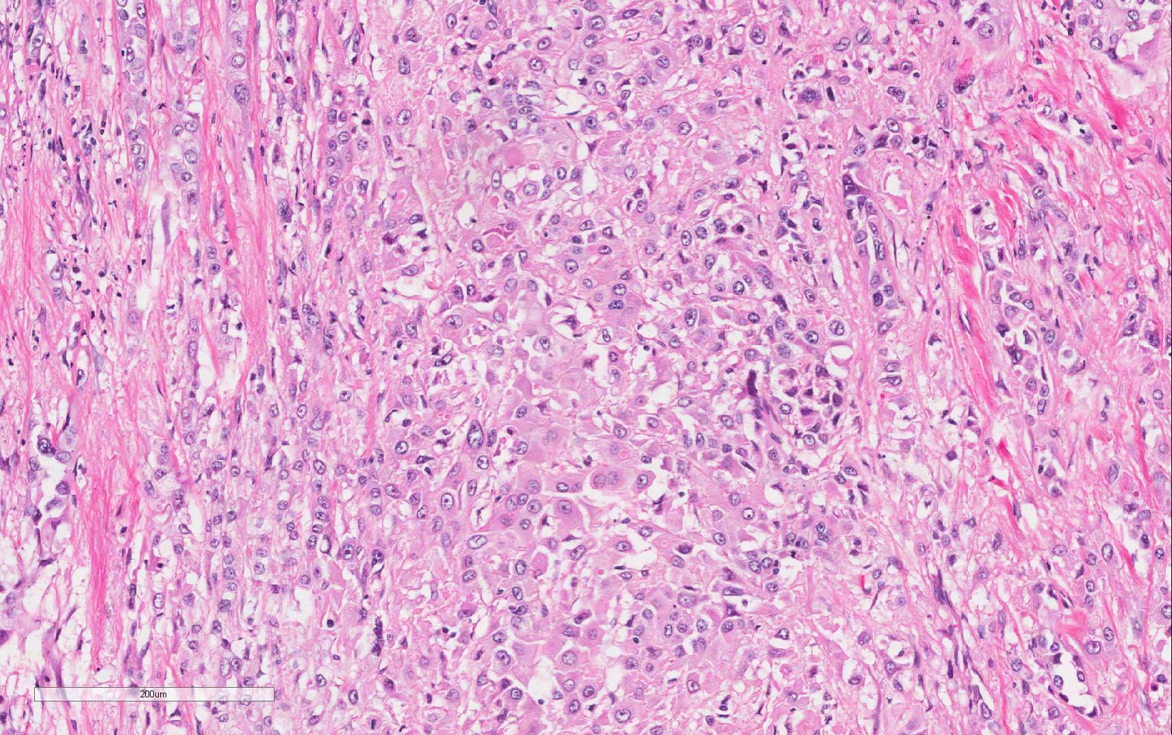
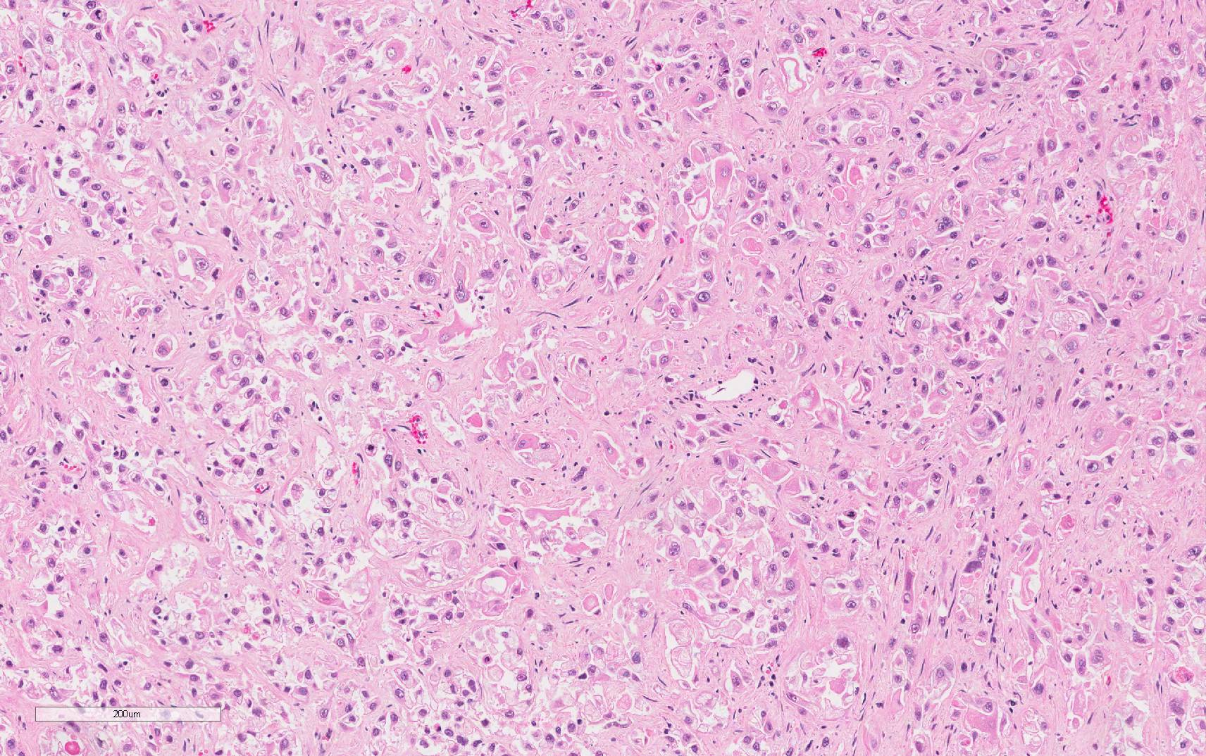
Microscopic Photographs:
 |
 |
|
| Left upper Lobe of Lung | Bone (Vertebra) | |
 |
 |
|
| Honey Combed Area | Omental Nodule |

 Meet our Residency Program Director
