Residency Program - Case of the Month
November 2013 - Presented by Jessica Rogers, M.D.
Clinical history:
A 70-year-old woman with malaise, hot flashes, and 5-10 pound weight loss was found to have an elevated hematocrit of 46. Additional labs showed a creatinine of 1.3, calcium of 10.8, an elevated transferrin and ferritin, a low erythropoietin level, negative for a JAK2 mutation. Imaging work-up at an outside facility showed for a 7.7 x 8.1 cm right renal mass in addition to masses up to 3.3 cm in sized that are posterior to the vena cava. She was referred to UC Davis Urology for further evaluation. Right total nephrectomy showed the following specimen:
Gross description:
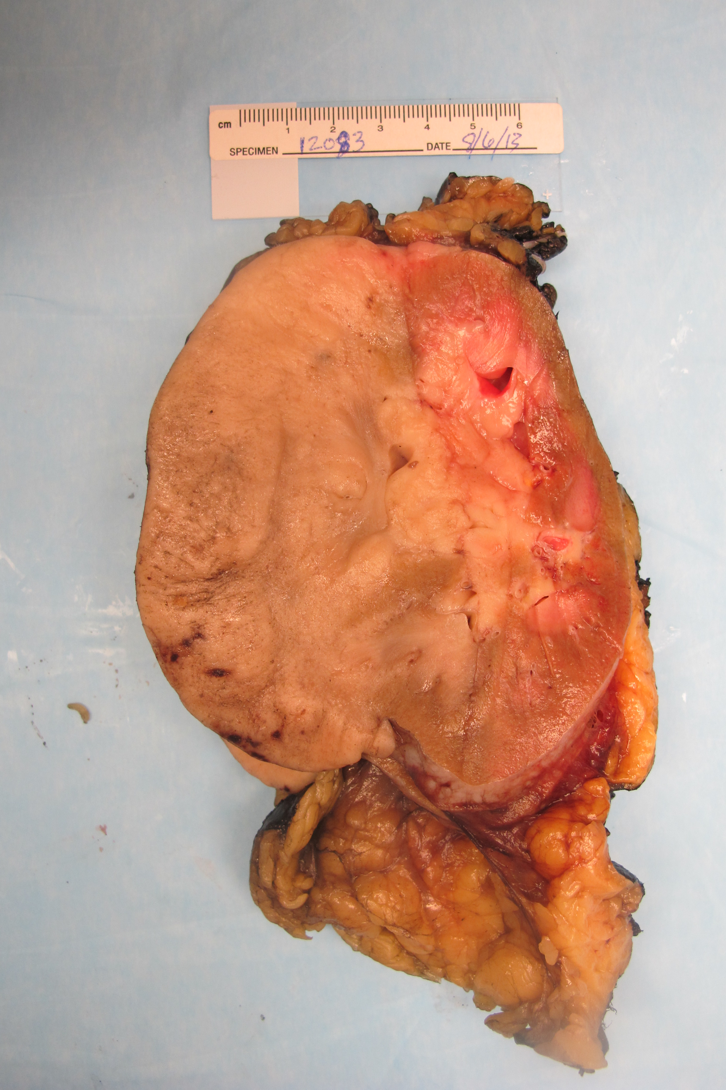
Received was a 590 gram kidney measuring 12.9 x 9.0 x 7.5 cm. Bivalving of the kidney revealed a light tan to tan-pink, solid tumor (11.7 x 5.8 x 6.8 cm) replacing the lateral aspect of the kidney. The tumor appears to be confined by the renal capsule (0.1 cm from the closest margin). At its interface with the renal parenchyma, sinus fat and collecting system the tumor had an infiltrative appearance, and grossly appeared to involve the sinus fat and calyces.
Micro description:
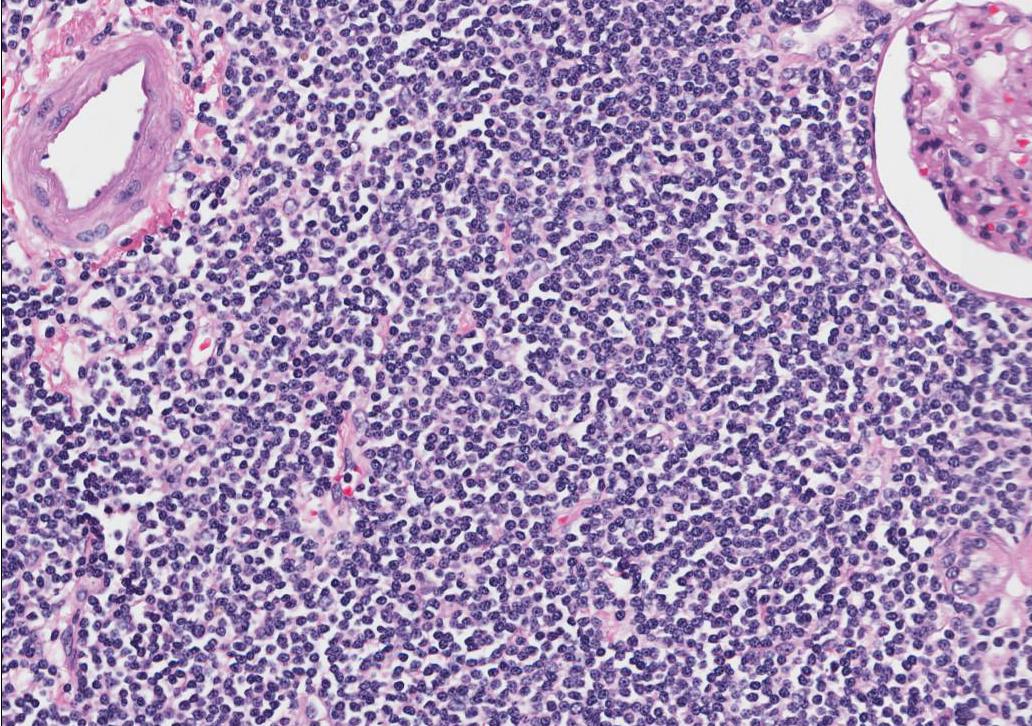
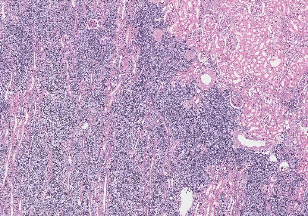
H&E stained sections of the kidney tumor and the paracaval lymph node demonstrated a diffuse, mature, monomorphic, atypical lymphoid proliferation and effacement of the normal organ and lymph node architecture. Most of the cells had minimal amount of pale cytoplasm, irregular nuclei and vesicular chromatin, rendering a mature lymphoid appearance. No sheets or aggregates of large cells were seen. Mitotic figures and apoptotic cells were difficult to find.
Immunohistochemistry:
|
Ki67 |
Low proliferative index. |
|
CD20 |
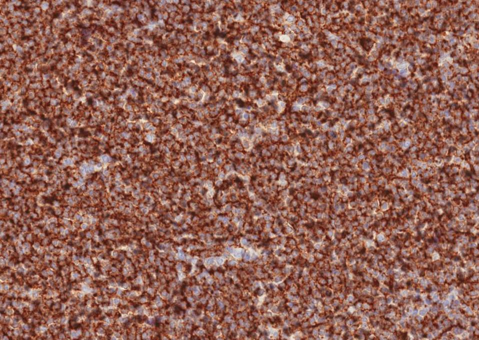
Highlights tumor cells and B cells. |
|
CD3 |
Highlights T cells. |
|
CD5 |
Negative in tumor cells. Highlights T cells. |
|
CD10 |
Negative in tumor cells. Highlights B cells. |
|
CD23 |
Negative in tumor cells. Highlights B cells. |
|
Cyclin D1 |
Negative. |
|
CD43 |
Partially positive in tumor cells. |
Gross photo:
Microscopic photos:
 |
 |
 |
||
| CD 20 | HE 200X | HE Low Power |


 Meet our Residency Program Director
