A priceless view
EyePod lets researchers see ‘what’s really happening’ inside tumors
Cancer is not like other diseases. It can grow rapidly and invade surrounding tissues, changing its molecular structure to evade treatments.
Because the disease is so dynamic, any single view — from a scan, biopsy or microscope — is just one frame in a long-running movie. To truly understand how cancer grows and adapts, we need to see the whole movie.
This plasticity has challenged researchers for decades. Scientists have tried various techniques to study how cancer develops over time, with mixed success. Cancer cell lines in a dish can adjust to their environment, adapting to the medium in which they grow. Biopsies are invasive, can only be performed infrequently, and they remove tumors from their native environments. Other approaches also can skew tumor behavior.
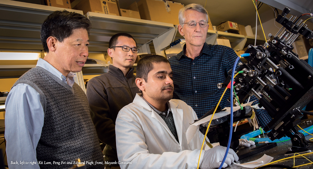
But now researchers at UC Davis have a new tool to follow cancer from its earliest, cellular stage. Using an optical imaging technology called the EyePod, which takes non-invasive, microscopic images of living mouse eyes, physiologist Edward N. Pugh, Jr., and cancer researcher Kit Lam are monitoring growth of human tumors implanted in mouse eyes in real time.
With this technology, scientists can watch how tumors grow, invade nearby tissues and recruit blood vessels, and observe the migration of host immune and vascular cells to the tumor site. Equally important, researchers can monitor experimental treatments, studying where they travel inside a tumor, where they may fall short, and how the tumor and host cells respond to treatments. As it turns out, the eye may be the window to cancer.
Building the EyePod
A professor in the UC Davis Departments of Physiology and Membrane Biology, and Cell Biology and Human Anatomy, Pugh has spent decades trying to understand how photoreceptors in the eye convert light into brain signals. His work, and that of other researchers, has shown how amazingly sensitive eyes can be.
“Rod photoreceptors can respond to a single photon of light,” notes Pugh. “These cells have achieved the ultimate sensitivity permitted by physics.”
But there was much more to learn from the eye, and Pugh needed new tools to get there. With funds from the UC Davis Research Investments in Science and Engineering (RISE) program, and in collaboration with Robert Zawadzki, assistant research professor in the Department of Ophthalmology and Vision Sciences, Pugh built the EyePod laboratory.
The idea was simple: create a suite of devices that use the mouse eye as a window to view cells in the neural retina, which is part of the central nervous system. By taking advantage of the eye’s natural optics, the researchers can image cells as if they were using a 20X microscope.
“We can now visualize individual cells and have imaged a single neuron for eight months,” says Pugh. “But equally important, we can image the entire capillary bed of the eye’s vasculature.”
This approach is completely non-invasive — it simply uses the mouse eye’s natural optics as a lens to see into the retina, which is a huge advantage. Optical techniques used to view the central nervous system or other internal tissues often require surgical or other invasive approaches, which can alter tissues and invalidate results. With the EyePod, researchers can observe without interfering with the normal biology.
A shared resource
The ability to record cell behavior inside the body offers incredible possibilities. UC Davis researchers are using the EyePod for various collaborative investigations, including the study of the immune system’s role in retinal degeneration and stem cell and gene therapy treatments for glaucoma and other diseases.
But it soon became clear to EyePod investigators that their technology was useful for more than the study of eyes and their specific diseases. The EyePod could be a powerful tool to examine a disease caused by malfunctioning cells.
Enter Kit Lam, who chairs the UC Davis Department of Biochemistry and Molecular Medicine, and is a co-leader of the cancer center’s Cancer Therapeutics program. He also recognized the EyePod’s potential to track cancer development. Long-running observations and cellular-resolution images could provide step-by-step insights into the devious ways tumors grow and ultimately lead to development of new and better therapies.
“The eye is a natural window that we can peek through and see over time: every hour, every day, every week, every month, even over years,” says Lam. “Through implanting tumor cells behind the retina of mouse eyes, we can understand how tumors develop, metastasize, form blood vessels and alter their microenvironment. If we treat the mouse with an anti-angiogenesis drug, we can see what happens to the blood vessels and the tumor. If we treat the mouse with immunotherapy, we can see how the host’s immune cells interact with the tumor before and after treatment.”
Developing new treatments
The EyePod also can help Lam perfect a technology his lab has been working on for several years — therapeutic nanoparticles.
One of the problems with chemotherapy is that it’s systemic. Chemo hits tissues throughout the body, both healthy and malignant.
However, Lam’s nanoparticles, which are 20 to 50 nanometers (one billionth of a meter) in diameter, can bring chemotherapy straight to the cancer. Because tumors often have leaky blood vessels, these nanoparticles are preferentially released inside them. Lam’s group also can attach peptides (pieces of proteins) that home in on specific proteins on the surface of the cancer cell.
Encased in these nanoparticles, treatments like doxorubicin pose little threat to normal tissue. But once they hit the tumor, the particles release their therapeutic payload, becoming anti-cancer smart bombs.
“The nanoparticle is a way to encapsulate this toxic drug and deliver it to the tumor site so the tumor gets more of the drug than does the normal tissue,” says Lam. “There will be less toxicity and more drug going to the tumor.”
With these nanoparticles, oncologists could deliver higher chemotherapy doses, which could increase treatment effectiveness. In addition, lowering toxicity could help patients who have trouble tolerating chemotherapy. When side effects become too harsh, these patients often must suspend therapy for a while to recover. Nanoparticles might provide more consistent treatment.
But developing this high-tech delivery system has been tricky. Researchers lacked important details about how nanoparticles function inside tumors. The EyePod gives Lam’s team the ability to actually watch these nanoparticles at work and find ways to improve them.
“By implanting tumor cells in the back of the eye, we can follow tumor formation and, more importantly, we can use the instrument to see how the nanoparticles distribute within the tumor in real time and how the tumor responds to treatment,” says Lam.
National significance
Combining the EyePod’s imaging abilities with nanoparticles has generated a lot of excitement. Last year the National Cancer Institute approved a five-year, $3 million grant to advance the project.
The team already has begun studying two of the most deadly forms of cancer: glioblastoma and non-small cell lung cancer. The hope is that new information from this research will improve patient care.
“This is a very fruitful collaboration — two different disciplines coming together to solve important problems,” says Lam. “With this technology, we are no longer shooting in the dark; we can see what’s really happening.”
