Challenging glioblastoma
New tools, models and targets against brain tumors
Medicine has made incredible progress against many cancers — but not glioblastoma multiforme (GBM).
The most common brain tumor, GBM is also the most aggressive. Patients rarely survive more than 18 months.
GBMs are difficult to treat for many reasons. Because these tumors often develop in sensitive brain regions, surgical and radiation oncologists must tread carefully. In addition, the blood-brain barrier, which protects us from pathogens and other invaders, blocks most chemotherapies.
Beyond that, GBMs are difficult to dislodge. Tumor tendrils penetrate the brain. Removing them has been likened to taking a wet spiderweb off a bush — microscopic remnants often remain.
These challenges have stumped the medical community for decades, but new approaches have potential to make great headway. Researchers and surgeons at UC Davis Comprehensive Cancer Center are studying new ways to control GBM. By hitting the disease from multiple angles, they hope to extend patient survival.
Better surgical margins
Surgical oncologists always want to remove as much cancer as possible, regardless of where the tumor is located, but working in the brain is especially challenging. Remove too little and the cancer comes roaring back. Remove too much and the patient can suffer cognitive damage.
Laura Marcu, professor of bio-medical engineering and neurological surgery, is developing technology that could help surgeons precisely remove GBMs without harming healthy tissue.
“It’s very hard to visually differentiate between cancer and normal brain tissue,” says Marcu. “The idea is to be able to distinguish, in real time, the brain regions where tumor cells are present from healthy brain tissue.”
Marcu’s team is solving this problem with light, or more specifically, the ways different tissues respond to light. She has spent several years developing a technology called fluorescence lifetime imaging (FLIM), which uses light to identify molecules in tissues.
“We’re taking advantage of the optical properties of different tissue types,” says Marcu. “Light carries information about the molecular makeup of tissue, and each molecule has a different signature. By measuring how long molecules emit light after they are excited by a low-energy laser beam, we can identify the type of tissue.”
In practice, a neurosurgeon deploys a handheld fiber optic probe to manually scan the surface of the brain or the cavity after a tumor has been removed. A viewer would instantly display information about each tissue’s fluorescence, helping the surgeon quickly separate cancer cells from normal ones.
This approach can augment presurgical MRI scans. Because the brain, and thus the tumor, often shifts during surgery, initial MRI scans become less useful. Real-time FLIM could eliminate that downside, allowing surgeons to reorient themselves during the procedure. In addition, the device requires no contrast agents or other markers; it simply sends light to the tissue and interprets the results.
This technology is being tested on patients but is still being developed. Eventually, a device company will have to step in to commercialize it. However, early results have been quite promising.
“We’re optimistic that we can distinguish the margins of the tumor using this label-free real-time approach,” says Marcu.
Cage the tumor
There are three main therapies associated with cancer treatment — surgery, chemotherapy and radiation. But now, a fourth technique is being used on GBM — electric fields.
“By generating an electrical field in and around a tumor, and by rapidly alternating that field, we can disrupt the normal process cells undergo for replication,” says Associate Professor and neurosurgeon Kiarash Shahlaie. “The therapy is not a cure, but it does buy more time.”
These alternating electric fields, called TTFields, can prevent cell division and even drive programmed cell death (apoptosis). TTFields are particularly well-suited for the brain, since neurons do not divide, and could eliminate cancer cells that remain after surgery.
The FDA has approved a TTFields instrument that uses electrodes on the scalp to generate the field. Clinical studies have shown the device increases survival, but there are drawbacks. Scalp electrodes can generate heat and other issues. In addition, because the field must penetrate both scalp and skull, it requires more power, which is supplied by a large external battery pack.
Patients had best results when they wore the device for 18 hours or more, but the discomfort, inconvenience and poor aesthetic sometimes discouraged compliance. People didn’t necessarily want to wear their electrical cap every day for the rest of their lives, regardless of the survival benefit.
Shahlaie and colleagues want to overcome these pitfalls with a miniaturized TTFields device. They are retasking technology used to implant electrodes inside the skull, similar to devices that treat epilepsy, Parkinson’s disease and other conditions. Wires would lead to a power source implanted in the chest, much like a pacemaker. Because the electrodes would be in the brain, they would require less juice to achieve the desired effect.
“My vision for this is a device that is permanently implanted,” says Shahlaie. “We could put electrodes in and around the brain to create an electrical cage around the tumor.”
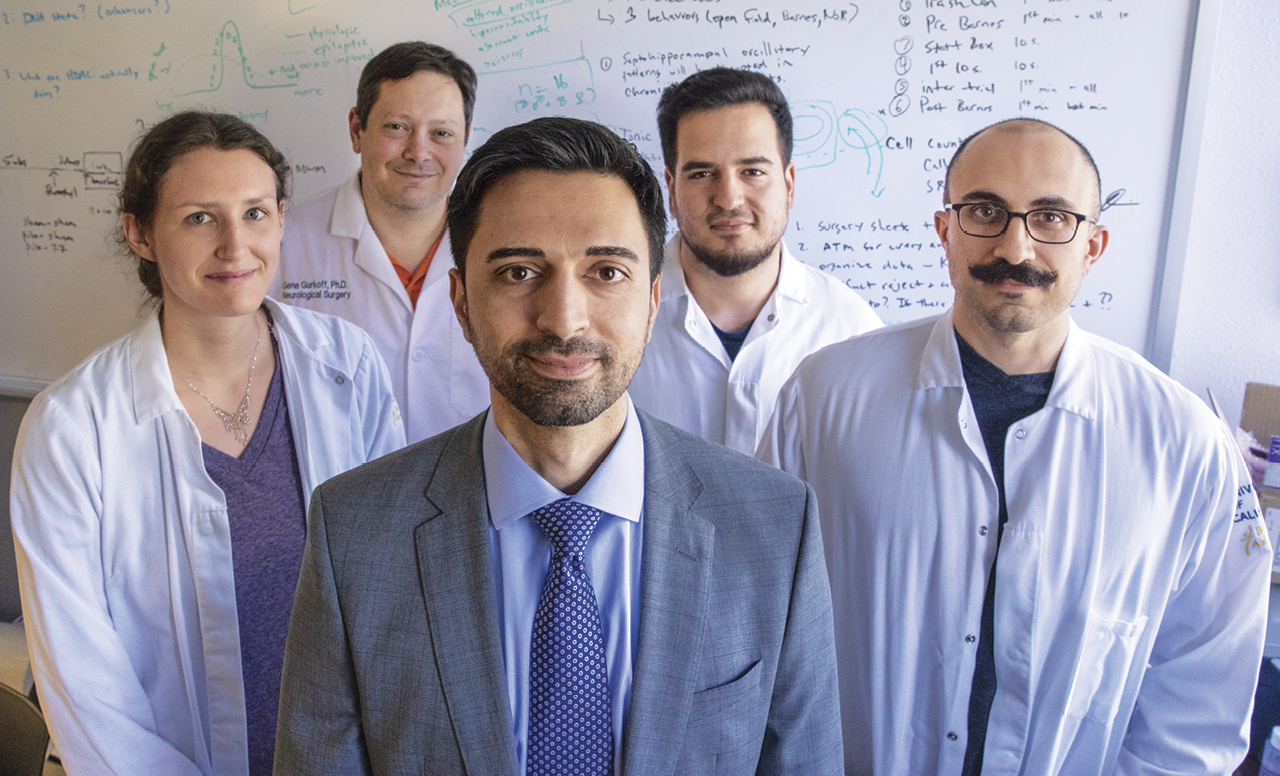
Shahlaie’s colleague Gene Gurkoff, a research scientist in the Department of Neurosurgery, said that will mean a real sense of freedom for patients.
“Having a fully implanted device means you could go to the hills and go camping, hiking or swimming and never have to worry about it,” he says. “You just turn it on and let it do its job.”
A nice target
Researchers are constantly looking for selective therapeutic targets — proteins and other molecules that play key roles in cancer but are not vital for healthy cells. One of these is a protein called ATF5, which is overexpressed in GBM and other cancers.
ATF5 is a transcription factor, a protein that helps turn on multiple genes. Targeting transcription factors can be difficult, because they interact directly with DNA and are often well-protected inside the nucleus. For many years, researchers felt most transcription factors were “undruggable.”
When James Angelastro was at Columbia University, he began investigating what would happen if he blocked ATF5. He created an ATF5 derivative — an engineered version of the protein that inhibits the original molecule’s function.
“I wanted to see if it caused cancer cells to differentiate,” said Angelastro, who is now associate professor in the Department of Molecular Biosciences at UC Davis. “Instead, I found that it killed cancer but not normal cells.”
Taking out ATF5 may destroy tumors because this protein regulates genes associated with cell survival. This is a big deal for cancer cells, which are often so damaged they need to turn up pro-survival mechanisms to simply exist. These survival genes also help tumors resist chemotherapy and radiation, but without them, cancer cells basically commit suicide.
The team has continued to tinker with their ATF5 molecule, adding a cell-penetrating peptide (a piece of a peptide) that helps it penetrate cancer cells. Angelastro’s ATF5 derivative penetrates the blood-brain barrier, a big hurdle for many treatments. The lab has tested the molecule in cell lines and animal models, and each time it destroyed GBM tumors. The molecule could also be effective in ovarian, breast, pancreatic, prostate, lung and other cancers with high ATF5 levels.
The technology has been licensed to Sapience Therapeutics, which is continuing the preclinical studies. UC Davis has an inter-institutional agreement with Columbia University, which holds the Sapience Therapeutics license.
“This is a great proof of concept that we can indeed target transcription factors,” says Angelastro. “This opens the door to new approaches to modulate ATF5 and perhaps similar molecules to eradicate cancer.”


